|
|
 |
Back to Annual Meeting Program
Laser Assisted Indocyanine Green Dye Angiography Accurately Predicts the Split-Thickness Graft Timing of Integra Artificial Dermis
Mitchell S. Fourman, M.Phil, Brett T. Phillips, MD, MBA, Jason R. Fritz, MS, Nicole Conkling, BS, Steve A. McClain, MD, Marcia Simon, Ph.D, Alexander B. Dagum, MD.
Stony Brook University Medical Center, Stony Brook, NY, USA.
BACKGROUND:
When addressing large surface area wounds, it is often not possible to use the patient's own skin as a grafting medium. Thus an artificial matrix such as Integra - a bilaminate combination of bovine tendon collagen and chondroitin-6-sulfate - is employed. Graft incorporation is a staged process beginning with fibroblast migration into the matrix, followed by endothelial cell mediated neovascularization by the second week of healing. Full incorporation is observed at the end of four weeks. Despite the advantages of Integra, complications have been reported relating to slow graft take or graft loss. Thus, it is of interest to determine the utility of Laser Doppler Imaging (LDI) and Laser Assisted Indocyanine Green Dye Angiography (LAICGA) in the tracking and prediction of Integra graft incorporation and split-thickness skin graft timing.
METHODS:
Two paraspinal full thickness wounds measuring 5x10cm were created on three female swine. Wounds were randomly assigned FTSG or Integra (Plainsboro, NJ) treatment, and both types of graft were placed following the application of Fibrin glue (Tisseel, Deerfield, IL) to the wound bed. LDI (Moor), LAICGA (LifeCell SPY) and clinical scoring were performed weekly for a period of 8 weeks post-graft. At six weeks the silicone layer of the Integra was removed, and a culture of autologous keratinocytes was applied. A 4mm punch biopsy was taken 1, 2, 4, 6, 7 and 8 weeks post-op for histology.
RESULTS:
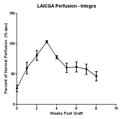
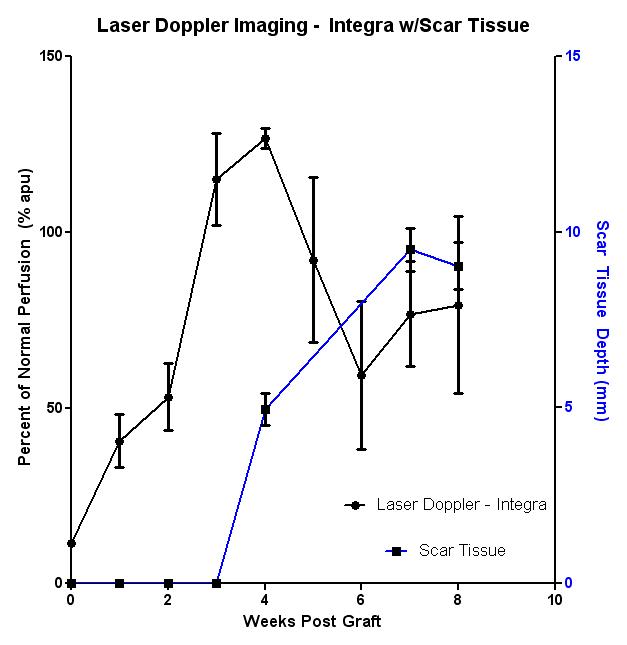
Both LAICGA and LDI perfusion measurements noted an increase in perfusion at the Integra graft site that peaked 3 weeks post-graft, corresponding with neovascularization timepoints noted in This peak perfusion corresponds with the timepoint at which split-thickness skin graft application would be appropriate. Perfusion settled to normal levels by 5 weeks post-graft. No significant perfusion changes were associated with autologous keratinocyte application. LAICGA measurements exhibit substantially greater regularity between animals at late timepoints as compared to LDI. This decrease in LDI resolution is directly related to increases in scar tissue above 5mm as determined via histologic analysis. This corresponds with what’s typically accepted as the maximum penetration depth of LDI signal.
CONCLUSIONS:
LAICGA perfusion measurements of Integra artificial matrix can serve as a valuable indicator of split-thickness graft timing. Further, LAICGA may provide valuable information as to graft integrity at late timepoints. LDI range appears to be insufficient for split-thickness graft timing or late timepoint accuracy. Future exploration of LAICGA potential will involve LAICGA tracking of Integra graft-delay in porcine models.
 
Back to Annual Meeting Program
|