|
|
 |
Back to Annual Meeting
Targeted cranial bone transport distraction is enhanced using a novel device
Mikell M. Yuhasz, BA, James Clune, MD, DMD, Felix P. Koch, MD, DMD, Rob Travieso, BS, Kenneth Wong, BS, Zhen W. Zhuang, MD, MS, Derek Steinbacher, MD, DMD.
Yale University School of Medicine, New Haven, CT, USA.
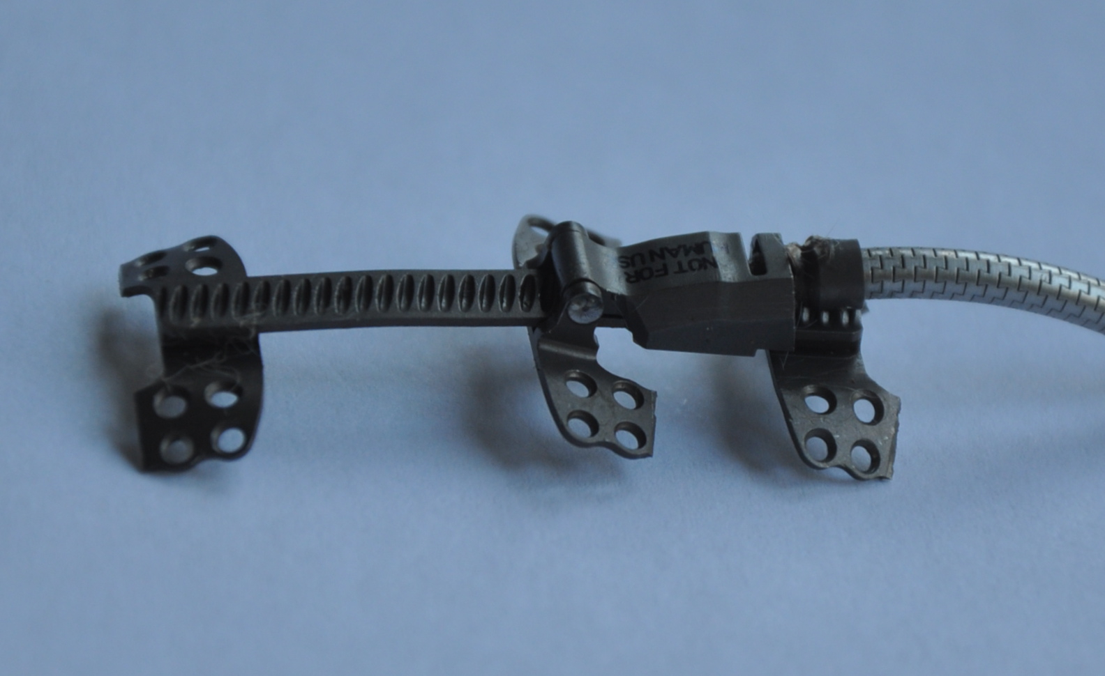
Background: Reconstruction of calvarial defects is challenging. Bone transport distraction osteogenesis (DO) has been described and carries the advantage of maintaining the osseous blood-supply and promoting slow soft tissue accommodation. The purpose of this study is to reconstruct a critically-sized rabbit calvarial defect using transport distraction via a novel, three-footplate device (Figure 1). We postulate that this targeted device allows for improved transport control, and requisite creation of contour.
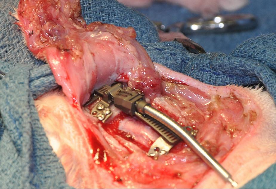
Methods: This study was approved by the Yale University IACUC (# 2011-11393). Four male New Zealand white rabbits (3 treatment and 1 control), aged 3 months were used. Following standard anesthesia, an anteriorly based, subperiosteal scalp flap was raised and a critical sized defect (16x16mm) created. An anteriorly abutting 10x16mm bone transport fragment was created in treatment animals. The distraction device was oriented anterioposteriorly, footplates secured posterior and anterior to the defect and segment, respectively, and a third footplate secured to the transport segment (Figure 2). The distraction protocol entailed one day of latency with subsequent activation of 1.5 mm per day. Consolidation occurred for 4 weeks post distraction. Following consolidation, animals were sacrificed and analyzed anatomically and histologically
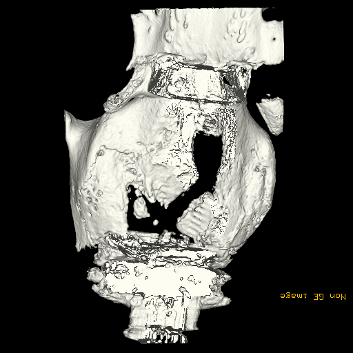
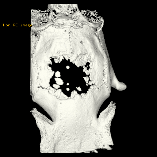
Results: There were no intra- or postoperative complications. The distraction protocol was well-tolerated. Gross inspection demonstrated regenerate bone in each treatment animal. Micro-CT demonstrated interposed mineralized regenerate bone between the distracted segments. The control animal failed to show significant ossification within the defect. Bone density measured >10.5% (range 7.8-12.4%) in the distracted group (Figure 3) versus 1% in the control (range 0.5-1.2%) (Figure 4). Histology revealed near uniform ossification and trabeculae between the distracted segments.
Conclusions: We demonstrate effective reconstruction of critical-sized rabbit calvarial defects using a novel transport distraction device. This modality enhanced targeted segment control and restitution of three-dimensional contour.
Back to Annual Meeting
|


