|
Back to Annual Meeting
Biomechanics of the thumb metacarpophalangeal joint in athletes: development of a computational model for injury prevention.
Arriyan S. Dowlatshahi, MD1, David A. Polito2, Joseph B. Rucco2, Tyler Wood2, Douglas M. Rothkopf, MD1.
1University of Massachusetts Medical School, Worcester, MA, USA, 2Worcester Polytechnic Institute, Worcester, MA, USA.
BACKGROUND
The thumb plays an eminent role in hand function. In particular, the metacarpophalangeal (MP) joint represents the necessary link between the mobile saddle joint and the finger itself, with requirements of stability and mobility. Injury is common in ballsports. Mechanisms of injury are poorly understood, and have disastrous consequences for hand function. In this study, a biomechanically accurate three-dimensional model of the thumb MP joint was created to allow for simulation and better understanding of prevalent injury patterns in clinical practice.
METHODS
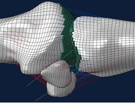
The finite element method has been successfully used in prior studies to accurately and precisely analyze the biomechanics of joints such as the hip, jaw, knee, and wrist. This type of analysis allows for the development of a computer-based model of physiological systems that can be subjected to various forces and displacements. (Fig. 1) Solid data was extracted from computed tomography images. The stabilizing soft tissues (capsular structures, abductor pollicis brevis, flexor pollicis brevis and adductor pollicis longus) were modeled using the finite element analysis software Abaqus™.
RESULTS
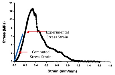
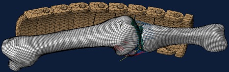
A three dimensional and biomechanically accurate thumb MP joint model was created. Using the software Abaqus™ it was subjected to forces of ulnar and radial deviation, flexion and extention. Stress-strain, stress-versus-time curves, shear-stress on the articular cartilage, forces experienced at ligament insertions were computed, and the biomechanical stabilizing function of the volar plate was examined. A particular focus was placed on the simulation of ulnar collateral ligament and hyperextention injuries. Internal validation of the model was obtained by comparing our computed ulnar collateral ligament measurements with previous results obtained by mechanical in-vitro testing. (Fig. 2) The applicability of our research is evidenced by the design of a protective device for the protection of the ulnar collateral ligament in soccer goalkeepers. (Fig. 3)
CONCLUSION
The finite element method was demonstrated to be a helpful tool to study the biomechanics of injury patterns involving the thumb MP joint. Practical applications include the conceptualization of protective devices, as well as their testing under extreme loading conditions which would otherwise not be possible in an in-vivo model. In addition, reconstructive efforts may be guided by the information gathered from this research.
Figure 1. Finite element model of the thumb metacarpophalangeal joint.
Figure 2. Stress strain curve of the ulnar collateral ligament of the thumb metacarpophalangeal joint.
Figure 3. Protective device designed to provide protection to the thumb MP joint.
Back to Annual Meeting
|


