|
|
 |
Back to Program Outline
Back to Posters
Topic:
Near-Infrared Imaging for Evaluation of Thrombosis in Microsurgery
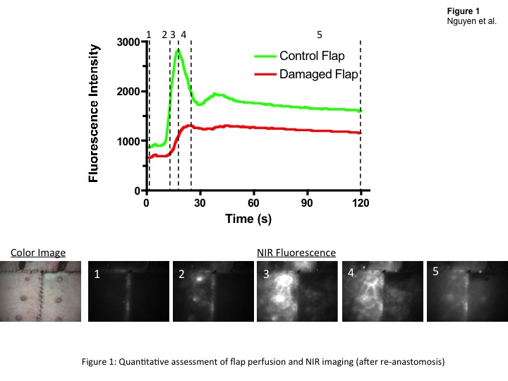
John T. Nguyen, MD, Yoshitomo Ashitate, MD, Vivek Venugopal, PhD, Florin Neacsu, MS, Frank Kettenring, BS, John V. Frangioni, MD PhD, Sylvain Gioux, PhD, Bernard T. Lee, MD, MBA.
BIDMC, Boston, MA, USA.
BACKGROUND:
Advances in techniques have increased the use of microsurgery over a wide range of clinical situations. Anastomotic site thrombosis, if not identified expeditiously, is a common cause of failure, resulting in flap loss. This study examines the use of intraoperative near-infrared (NIR) fluorescence imaging to identify microsurgical anastomotic patency, thrombus formation, and resolution.
METHODS:
Bilateral abdominal flaps were designed in Yorkshire pigs using the deep superior epigastric artery and vein pedicle. A validation study was performed in 12 flaps (n = 6 subjects) to develop a reproducible model for complete thrombosis. Once a reproducible thrombosis model was created, bilateral flaps (n = 4 subjects) were harvested and a micro-vascular anastomosis performed on one side as thrombosis was induced with ferrous chloride. This was compared to an intact flap on the contralateral side. NIR fluorescence imaging was performed after flap elevation, microsurgical anastomosis, thrombosis formation and resolution.
RESULTS:
Vessel patency after flap elevation, microsurgical anastomosis, and complete thrombosis were confirmed with NIR imaging. In a healthy case (no thrombosis) fluorescence intensity increased by 74% to 132% within 2 minutes. With application of ferrous chloride, NIR demonstrated lack of flow through the vessels and perforators with a fluorescence increase of only 13% within 2 minutes. After resolution of clot, the anastomosis was revised and flow was re-established through the pedicle with a fluorescence increase of 75% within 2 minutes (Figure 1). In addition, we also identify two distinct phases in the fluorescence intensity dynamic providing information regarding vascularization and perfusion, respectively.
CONCLUSIONS:
Intraoperative evaluation of micro-vascular anastomosis in free flaps is possible with a real-time, NIR fluorescence imaging system. Early studies with these imaging systems in our laboratory show promise in identifying thrombosis formation and impairment of blood flow with simultaneous assessment of tissue perfusion. 
Back to Program Outline
Back to Posters
|