|
|
|
|
|
Back to 2015 Annual Meeting
Hyperspectral imaging as method of skin flap necrosis prediction in a murine model
Ava G. Chappell, BA1, Michael S. Chin, MD1, Giorgio Giatsidis2, Dylan Perry, BA1, Jorge Lujan-Hernandez, MD1, Anthony Haddad, MD2, Hajime Matsumine, MD PhD2, Dennis P. Orgill, MD PhD2, Janice F. Lalikos, MD1.
1University of Massachusetts Medical School, Worcester, MA, USA, 2Brigham and Women's Hospital, Boston, MA, USA.
Background:
Necrosis remains a significant complication in cutaneous flap procedures. Objective methods such as fluorescence spectroscopy, laser Doppler velocimetry, intraoperative indocyanine green angiography and fluorescein dye administration have been investigated as tools to aid flap assessment. Despite these methods, there is still no clinical standard.
Objectives:
In this study, we investigate the use of hyperspectral imaging (HSI) to correlate early skin perfusion changes and ultimate flap survival in a murine model.
Methods:
Thirty-one hairless, immunocompetent, adult male mice were used. Random patterned dorsal skin flaps (1.5x3cm) were elevated and sutured back into place with an impermeable barrier. Hyperspectral and digital images were obtained 30 minutes after flap elevation, 24 hours, 72 hours and prior to sacrifice on post-op day 7.
Results:
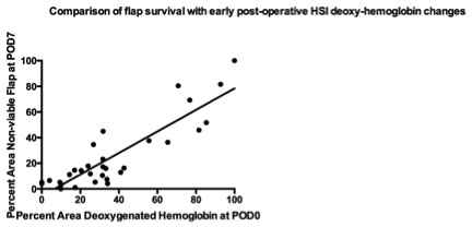
HSI data acquired from each mouse were retrieved to obtain the deoxygenated hemoglobin maps of the skin flap at 30 minutes post-op. Measurement of the corresponding area was performed of the visible demarcated area in digital photographs on post-op day 7. Pearson correlation demonstrated statistically significant relationship between percent area demarcated by deoxygenated hemoglobin at 30 min and clinically necrotic area seen on sacrifice day (r^2=0.4848, p <0.0001).
Conclusion:
These findings suggest that early changes in deoxygenated hemoglobin seen with HSI may predict the region and extent of flap necrosis. Since HSI is non-invasive, user-friendly technology, this could be a useful tool to help providers monitor skin flap survival in the clinical setting.
Back to 2015 Annual Meeting
|
|


