Back to 2015 Annual Meeting
Neonatal Mandibular Distraction Osteogenesis: Virtual Surgical Planning Becomes Operative Reality
Matthew Doscher, MD1, Jillian E. Schreiber, BA2, Carrie Stern, MD1, Evan Garfein, MD1, James Goodrich, MD, PhD1, Oren Tepper, MD1.
1Montefiore Medical Center, Bronx, NY, USA, 2Albert Einstein College of Medicine, Bronx, NY, USA.
PURPOSE: Mandibular distraction osteogenesis (DO) has become an accepted method to manage severe cases of micrognathia-induced airway obstruction in neonates. Current imaging offers only a rough guide for operative planning. This leaves a significant obstacle to translating the surgical plan. To our knowledge, we offer the first description of computerized virtual surgical planning used to help guide pre- and intra-operative steps for neonatal mandibular DO.
METHODS: A virtual surgical plan was created and tested using three-dimensional reconstructions of the patients' CT scans, custom guides and distraction devices. The plan simulated the positioning of the osteotomy, device and distraction vector. In the operating room the virtual plan served as a step-by-step guide for the execution of the procedure.
RESULTS: This unique approach to surgical planning of distraction osteogenesis was used for 5 neonates. As predicted from testing, each mandible was unique such that the customized guide would only "snap-on" if appropriately placed in the planned position. After securing the guide with K wires the osteotomy was performed. The guide and distraction device were exchanged by sliding over the K wires using the preplanned holes. The device was then secured. An average advancement of 18.5 mm was performed bilaterally. Hardware was removed 3 months post-operatively. At follow-up the children have excellent cosmetic results and have successfully avoided tracheotomy. Additionally, similar planning methods were used with 2 patients for Le Fort III distractions with successful results.
CONCLUSIONS: The custom guides created through 3D printing allowed for seamless transfer from virtual plan to operative steps. This provides objective guidance in device selection, vector planning and operative guide positioning both for mandible and midface distraction.
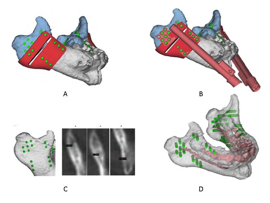
Figure 1 - (A) 3D virtual model of the patient's mandible with the cutting guide in place - green areas represent potential screw position. (B) Virtual model showing both the distraction device and cutting guide overlaid on one another. (C) For each of the preplanned screw positions, the thickness was measured to pre-plan the appropriate screw for a bicortical bite. (D) With virtual demonstration of the nerve position, screw position was identified to avoid the nerve.
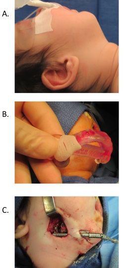
Figure 2 - The use of 3D printed cutting guides for placement of the distraction device are shown. (A) Micrognathic patient with Pierre Robin Sequence. (B) 3D model of the patient's hypoplastic mandible is fitted with the 3D-printed cutting guide. The 3D-printed cutting guide was then fitted on the patient's mandible to guide the osteotomy and screw position for distraction device placement (C) Placement of the distraction device on the patient's mandible.
Back to 2015 Annual Meeting
|


