|
|
|
|
|
Back to 2015 Annual Meeting
Are 2D Pictures Becoming "Faux-Tography?" the Value of Incorporating 3D Photography into Your Aesthetic Practice
Jillian E. Schreiber, BA1, Carrie Stern, MD2, Evan Garfein, MD2, Elizabeth Jelks, MD3, Glenn Jelks, MD3, Oren Tepper, MD2.
1Albert Einstein College of Medicine, Bronx, NY, USA, 2Montefiore Medical Center, Bronx, NY, USA, 3Jelks Medical, New York, NY, USA.
Background
Most aesthetic practices today rely on conventional 2D photography for patient assessment and surgical planning. 3D photography is used by a small percent of surgeons primarily to enhance the consultation with simulation. Yet, the value of 3D photography extends well beyond marketing with the potential to significantly enhance patient analysis and implementation of a surgical algorithm. The goal of this study was to assess the clinical utility of 3D photography and determine procedures that would most benefit from a 3D analysis report.
Methods
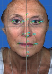
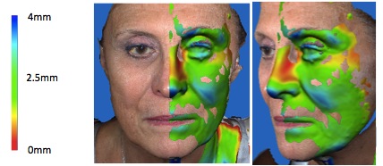
Since July 2014, the senior authors (OT, GW) have included 3D photographs as part of the pre-operative work-up for patients undergoing facial aesthetic procedures. 3D photographs were obtained with a hand-held camera (Canfield Vectra H1) or automated booth (Di3D System). A standardized pre-operative 3D report was generated that included: a mid-sagittal plane, placement of key landmarks, and calculation of surface and vector distances from the midline. (Figure 1) Each hemi-face was reflected to the opposite side such that two new composite 3D faces were created, termed right and left "hemi-composites". A symmetry report was then generated and color maps produced by calculating differences in volume and projection. (Figure 2)
Results
The dimensional accuracy of the photographs did not differ between the stationary and portable capture system, which required stitching based on selection of key landmarks. Of the various measurements provided in the 3D analysis report, ‘symmetry' analysis proved to be the most valuable component. During the preoperative consultation, composite views highlighted inherent asymmetry and helped patients identify "favorable" features. Symmetry reports and composite views provided a means of facial analysis, far superior to relative to standard 2D photos.
Color volume maps highlighted the region and severity of contour asymmetries. Fat transfer maps for targeted injection were generated, which was a significant improvement from traditional method of subjective patient markings. We found this particularly useful in lipostructure used to augment medial and lateral cheek compartments and for blending of the lid-cheek junction.
Conclusions
3D photography is valuable tool in facial aesthetic surgery with various applications. Symmetry analysis can supplement surgical simulation and help align patient preferences with surgeon goals. A standard 3D analysis report provides objective measurements of facial anatomy, which can help guide treatment. Ongoing studies are looking at 3D photography as an intra-operative reference, as well as post-operatively to measure surgical outcomes. Based on our experience, we believe 3D photography will play an increasing role in plastic surgery practices and ultimately may become standard of care for patient assessment and surgical planning.
Legend:
Figure 1. 3D pre-operative photograph with key landmarks and x,y,z coordinates.
Figure 2. Color map quantifying facial asymmetry. Demonstrates more severe malar descent and jowling of left hemiface (blue).
Back to 2015 Annual Meeting
|
|


