Back to 2016 Annual Meeting
Teaching an Old Dog New Tricks: The Composite Extended Paramedian Forehead Flap- A Cadaveric Study and Clinical Applications
Sabrina Pavri, M.D., Brandon Sumpio, B.A., Alain Kaldnay, B.A., Ajul Shah, M.D., Andrew W. McGregor, M.D., Flora Levin, M.D., Deepak Narayan, M.D..
Yale University, New Haven, CT, USA.
Introduction
The classic paramedian forehead flap is one of the most commonly used pedicled flaps for nasal reconstruction. Previous anatomical studies have found that the supratrochlear artery traverses the supraorbital rim and divides quickly into deep and superficial branches. It has been demonstrated that the periosteum and the frontalis-galea layer of the forehead can remain independently vascularized from an anterior (supraorbital/supratrochlear) pedicle as long as the dissection remains at least 10mm cephalad to the supraorbital rim.
We hypothesize that the periosteum and frontalis of the forehead have a blood supply originating from the supratrochlear artery that is independent of the overlying skin and subcutaneous tissue and can be recruited to enhance the more limited cutaneous paddle of the paramedian forehead flap.
Methods and Materials
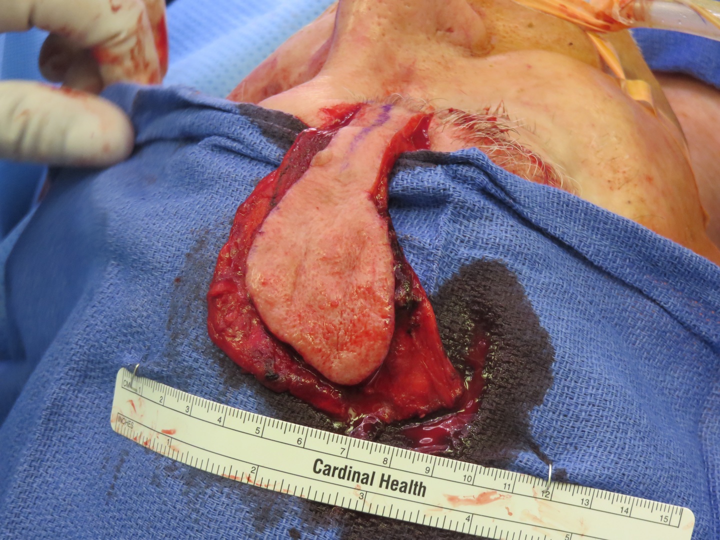
Six cadaver heads were used to analyze the vascularity of the composite extended paramedian forehead flap. In four of the cadaver heads, following flap dissection methelyene blue was injected into the internal carotid artery to confirm perfusion of the frontalis and periosteum outside of the cutaneous footprint (Figure 1/2). In the remaining two cadaver heads, following flap dissection the internal carotid artery was injected with a mixture of Omnipaque and gelatin and the heads were imaged with a microCT scan to confirm the vascular anatomy.
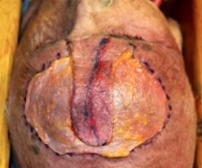
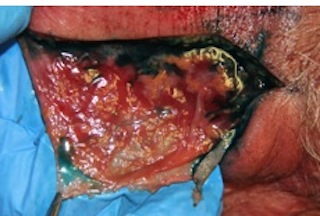
Three selected patients underwent the reconstructive use of the composite extended paramedian forehead flap for reconstruction of an orbital exenteration, a heminasal Mohs defect, and a total rhinectomy defect.
Results
Methylene blue injection confirmed vascular flow to the lateral frontalis and periosteum of the extended composite forehead flap. They will be evaluated by microCT to confirm our hypothesis on imaging studies.
The composite extended paramedian forehead flap has been used in three separate clinical cases with successful results. Intraoperative dissection of one of the flaps used is shown in Figure 3. The use of indocyanine green assisted imaging was used in two of the three cases to confirm viability of the extended lateral portion of the flaps. There was no evidence of any flap complications in the selected cases.
Conclusion
The composite extended paramedian forehead flap can be used successfully in single stage reconstruction of large nasal and orbital defects that would otherwise require several staged procedures or free tissue transfer.
Footnotes
1 Reece EM, Schaverian M, Rohrich RJ. “The Paramedian Forehead Flap: A Dynamic Anatomical Vascular Study Verifying Safety and Clinical Implications.” Plast. Reconstr. Surg. 121: 1956, 2008.
2 Yoshioka N, Rhoton Jr. AL. “Vascular Anatomy of the Anteriorly Based Pericranial Flap.” Neurosurgery 57[ONS Suppl 1]:ONS-11–ONS-16, 2005.
Back to 2016 Annual Meeting
|
