|
|
|
|
|
Back to 2016 Annual Meeting
Extracellular Matrix Impregnated with Adipose Derived Stem Cells in Skeletal Muscle Regeneration following Volumetric Muscle Loss in a Murine Model
Michael A. Plastini, M.D1, Shaohua Chang, PhD2, Nikolas Kappy, MD1, William Harris, MD1, Telisha Ortiz1, Ping Zhang, PhD3, Jeffery P. Carpenter, MD1, Spencer A. Brown, PhD3.
1Cooper University Hospital, Camden, NJ, USA, 2Cooper University Hospital Depatment of Surgical Research, Camden, NJ, USA, 3Cooper University Hospital Department of Surgical Research, Camden, NJ, USA.
BACKGROUND:
This study aims to address the need for a viable platform in soft tissue reconstruction, specifically following volumetric muscle loss (VML). We hypothesized that the extracellular matrix (ECM) stimulated muscle regenerative response demonstrated in previous studies1 would be further enhanced with an ECM impregnated with adult adipose derived stem cells (ASC) under optimized conditions.
METHODS:
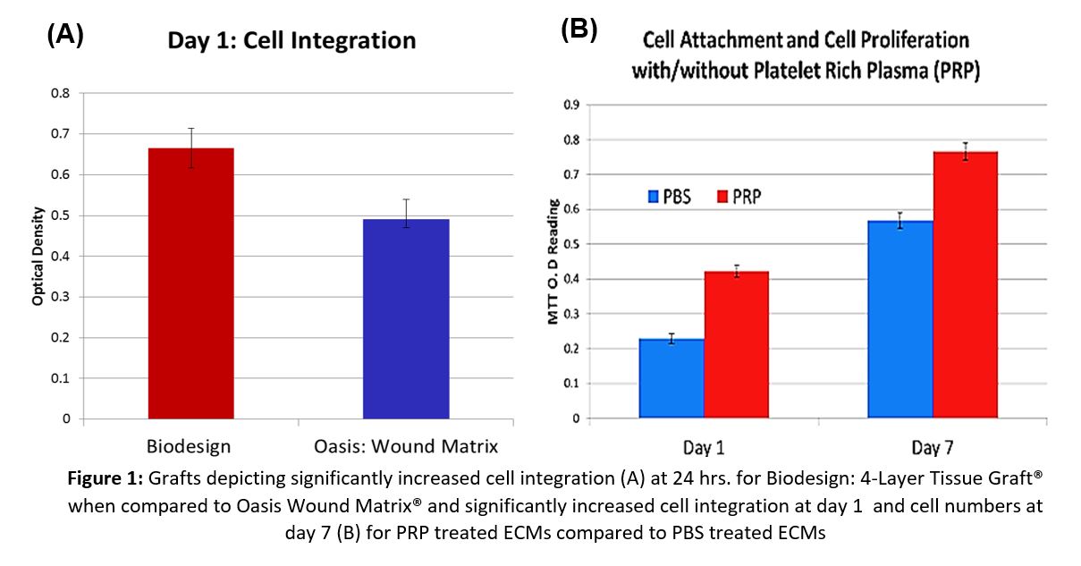
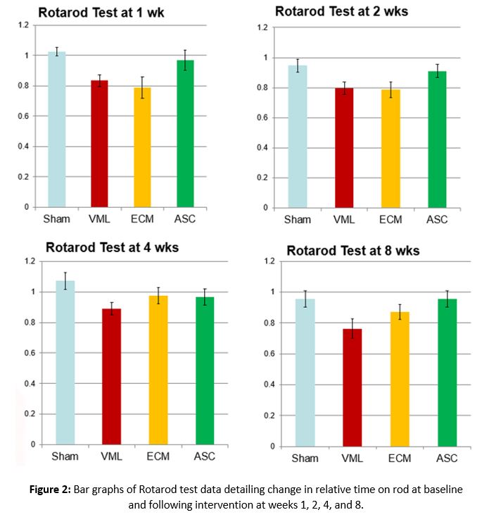
Biodesign: 4-Layer Tissue Graft® (BDTG) (n=4) and Oasis Wound Matrix® (OWM) (n=4) were compared. Each ECM was incubated in phosphate-buffered saline (PBS) solution or 5% platelet rich plasma (PRP) solution for 24 hours containing ASC maintained in a cell bank at our institution. MTT assays, cell growth curves, and light microscopy testing were utilized to assess cell attachment and proliferation. BDTG was then used in a VML murine model following resection of the tensor fasciae latae. Study groups included VML without repair (n=6), implantation of ECM without ASC following VML (n=6), and implantation of ECM impregnated with ASC seeded in a 5% PRP solution following VML (n=6). A sham group (n=6) was utilized for baseline testing. All mice underwent gait analysis at weeks one, two, and four utilizing Rotarod testing, and then were euthanized to examine histological and immunohistochemical evidence of muscle incorporation and regeneration.
RESULTS:
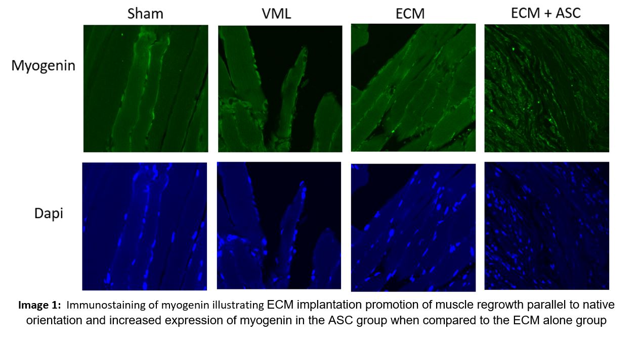
In vitro data demonstrated significantly increased ASC attachment (P<0.001) and a trend towards increased proliferation (P=0.243) for BDTG when compared to OWM. PRP solution significantly increased ASC attachment (P<0.001) and proliferation (P<0.001) on both ECMs when compared to PBS controls (Fig. 1). In vivo data at week one revealed relative time on Rotarod when compared to baseline were equivalent in the sham animals (1.02±0.03) but significantly decreased for VML alone (0.83±0.04), ECM (0.79±0.07), and ASC (0.97±0.07) mice (n=6, p=0.036) (Fig. 2). Myogenin staining (Image 1) showed: expected compensatory muscle hypertrophy in all VML animals; ECM implantation promoted muscle regrowth parallel to native orientation; and increased expression of myogenin in the ASC group when compared to the ECM alone group.
CONCLUSION:
We demonstrated that BDTG represents a superior platform when compared to OWM for ASC integration and proliferation. Also, the addition of ASC to ECM increased muscle regeneration and functional recovery in a VML model. We anticipate that the knowledge gained in this study will lead to better clinical outcomes in soft tissue reconstruction in future studies.
References:
1. Sicari, B.M., et al., An acellular biologic scaffold promotes skeletal muscle formation in mice and humans with volumetric muscle loss. Sci Transl Med, 2014. 6(234): p. 234ra58.
Back to 2016 Annual Meeting
|
|
