Back to 2016 Annual Meeting
Re-establishment of Lymphatic Drainage Patterns after Vascularized Lymph Node Transfer in a Rat Model
Marc Najjar, MD, Marcos M. Lopez, Jr., BS, Yelena Akelina, DVM, MS, Jeffrey A. Ascherman, MD.
Columbia University, New York, NY, USA.
BACKGROUND: Vascularized lymph node transfer (VLNT) has recently received attention as a potential surgical treatment for lymphedema. Despite good results in some series, the mechanism and benefits of VLNT have yet to be fully understood. In order to scientifically investigate the feasibility, viability and limitations of VLNT, we are using rats as an animal model. We have previously demonstrated the technical feasibility of VLNT in a rat, as well as the viability of transferred lymph nodes. The aim of the current study is to assess the re-establishment of drainage into transferred lymph nodes following VLNT, and to determine the time required for this to occur.
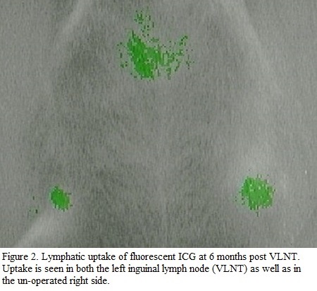
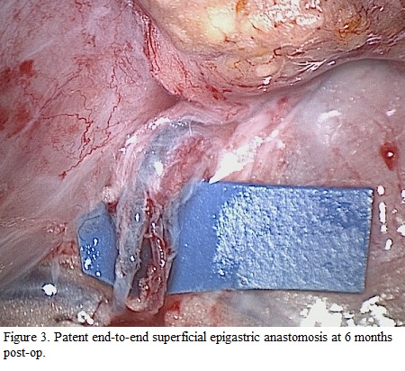
METHODS: Seven Sprague-Dawley rats were used to study the re-establishment of lymphatic flow into transplanted lymph nodes after inguinal VLNT. The operation performed on each rat consisted of two parts. First, the left groin lymph node basin with superficial epigastric vessels was harvested as a free flap. Second, the flap was re-attached in the left groin of the rat via end-to-end microvascular anastomoses of superficial epigastric vessels. Anastomosis patency was assessed visually immediately post-op and after 6 months. The rats were evaluated for re-establishment of lymphatic flow into the transplanted nodes at 1 month intervals up to 6 months post-op. This was accomplished non-invasively by injecting the rats in their flanks with fluorescent indocyanine green (ICG). ICG uptake was then detected using a PDE infrared camera. After six months, the rats were sacrificed to assess for anastomotic patency and lymph node viability by histology.
RESULTS: VLNT was performed in 7 rats. Anastomoses were patent in all rats immediately post-op. The rats were followed for 6 months to assess lymphatic re-establishment. No ICG uptake was seen in the transplanted lymph node basins in the first two months post-op (Figure 1). In 5 of 7 rats, ICG uptake was demonstrated in the transplanted lymph node at an average of 13 weeks, consistent with lymphatic channel re-establishment (Figure 2). So far, 5 rats have been sacrificed after 6 months and were found to have patent anastomoses (Figure 3). Histology results are currently pending, and will be included at the time of presentation.
CONCLUSIONS:
VLNT can be successfully performed in a rat model. We have previously shown feasibility and viability of VLNT in the lower extremity of rats. We now report uptake of ICG in 5 of 7 rats at an average of 13 weeks following transplantation, consistent with the re-establishment of lymphatic drainage into the transplanted lymph nodes after approximately 3 months.
Back to 2016 Annual Meeting
|
