|
|
|
|
|
Back to 2016 Annual Meeting
IL-10 Improves BMP-2-Mediated Bone Regeneration in the Infected Calvarial Defect in Rabbits
Michael R. Bykowski, M.D.1, Liliana Camison, M.D.1, Lee Weiss, Ph.D.2, Phil Campbell, Ph.D.2, Gregory M. Cooper, Ph.D.1, Joseph E. Losee, M.D.1.
1University of Pittsburgh Medical Center, Pittsburgh, PA, USA, 2Carnegie Mellon University, Pittsburgh, PA, USA.
Background: Complex craniofacial wounds resulting in calvarial defects are not only seen in the combat soldier but also in the civilian population. Contamination with multiple pathogens is common with these wounds and often results in indolent infection. Infection limits options for primary salvage and can impair secondary reconstruction by inhibiting bone healing, causing tissue scarring, and affecting overall functional recovery. In order to further improve this potential therapeutic modality, we are looking to novel strategies such as biopatterning of degradable matrices with biologics and cytokines to enable localized delivery of therapeutics. This strategy will simultaneously target both the immune and osteogenic responses at the site of wound repair or reconstruction. The purpose is to improve the treatment of craniofacial bone defects complicated by infection and scarring.
Methods: 16-week old New Zealand White rabbits underwent the standard 15mm x 15mm defect in the dorsal cranial vault defect. Following the defect, the removed bone flap was inoculated with S. aureus and immediately replanted into the defect. After an infection period of one week, the infected autograft was removed and the defect surgically debrided of devitalized tissue. After a two-week course of antibiotics, the animals were then underwent the final surgery with acellular dermal matrix (ADM) only (n=7), ADM + IL-10 (n=6), ADM + BMP-2 (n=8), or ADM + BMP-2/IL-10. All growth factors and cytokines were bioprinted on the ADM, as previously described. Following the last surgery, rabbits underwent CT scans at days 0, 14, 28, and 42 for evaluation of bone regeneration.
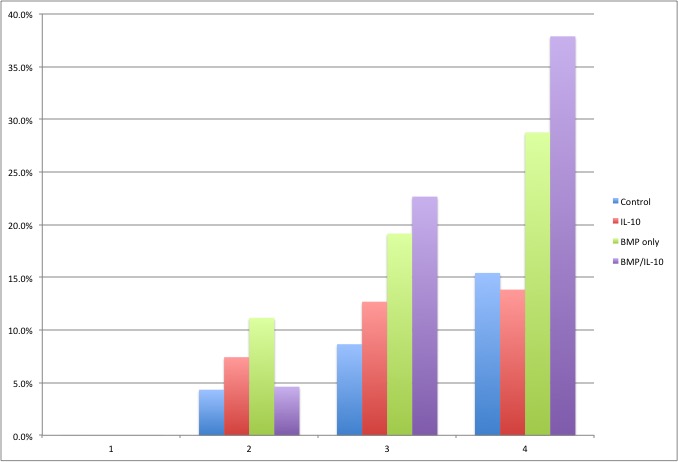
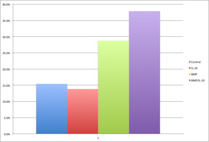
Results: In all experimental groups, the bone fill increased progressively over the 42-week period (see figure). At 42 weeks, the mean bone regeneration for each groups is as follows: ADM alone= 15.4%, ADM + IL-10= 13.8%, ADM + BMP-2= 28.8%, and ADM + BMP-2/IL-10= 37.9% (see figure). The BMP-2/IL-10 group had significantly more bone at 42 weeks compared to the control (ADM alone) and the ADM + IL-10 group (both p’s <0.03).
Conclusions: Calvarial defects are common in both the military and civilian populations. The bony defect is especially recalcitrant following infection, which is a potential complication following treatment for hemorrhagic strokes, trauma or cancer-related surgery. Our study demonstrates, in a clinically relevant craniofacial model, that bone regeneration can be improved in infected calvarial defects through growth factor therapy. To our knowledge, this is the first time that IL-10 has been shown to improve BMP-2-mediated bone regeneration.
Back to 2016 Annual Meeting
|
|
