Back to 2016 Annual Meeting
Validating Outcome Metrics: Accuracy of the VECTRA H1 Portable 3D Photogrammetry System for Facial Imaging Applications
Liliana Camison, MD, Michael Bykowski, MD, Wei Wei Lee, BS, Jesse A. Goldstein, MD, Joseph E. Losee, MD, Seth Weinberg, PhD.
University of Pittsburgh, Pittsburgh, PA, USA.
BACKGROUND: 3D imaging using stereophotogrammetry has become increasingly popular in plastic surgery and related fields, offering great advantages for surgical planning and outcome evaluation. Several commercial systems are currently available with varying features. In contrast to previous versions that require stationary set-up stations and previous calibration, the handheld VECTRA H1 is a low cost, highly portable system designed for facial imaging that is comparable to a common SLR camera. These attractive features have made the H1 very popular in clinical practice already. However, the H1 system has not been scientifically validated in independent studies, and the portability may come at a cost in terms of accuracy of surface registration. The purpose of this study was to validate the VECTRA H1 by comparing it to a throughly studied stationary system, the 3DMdFace.
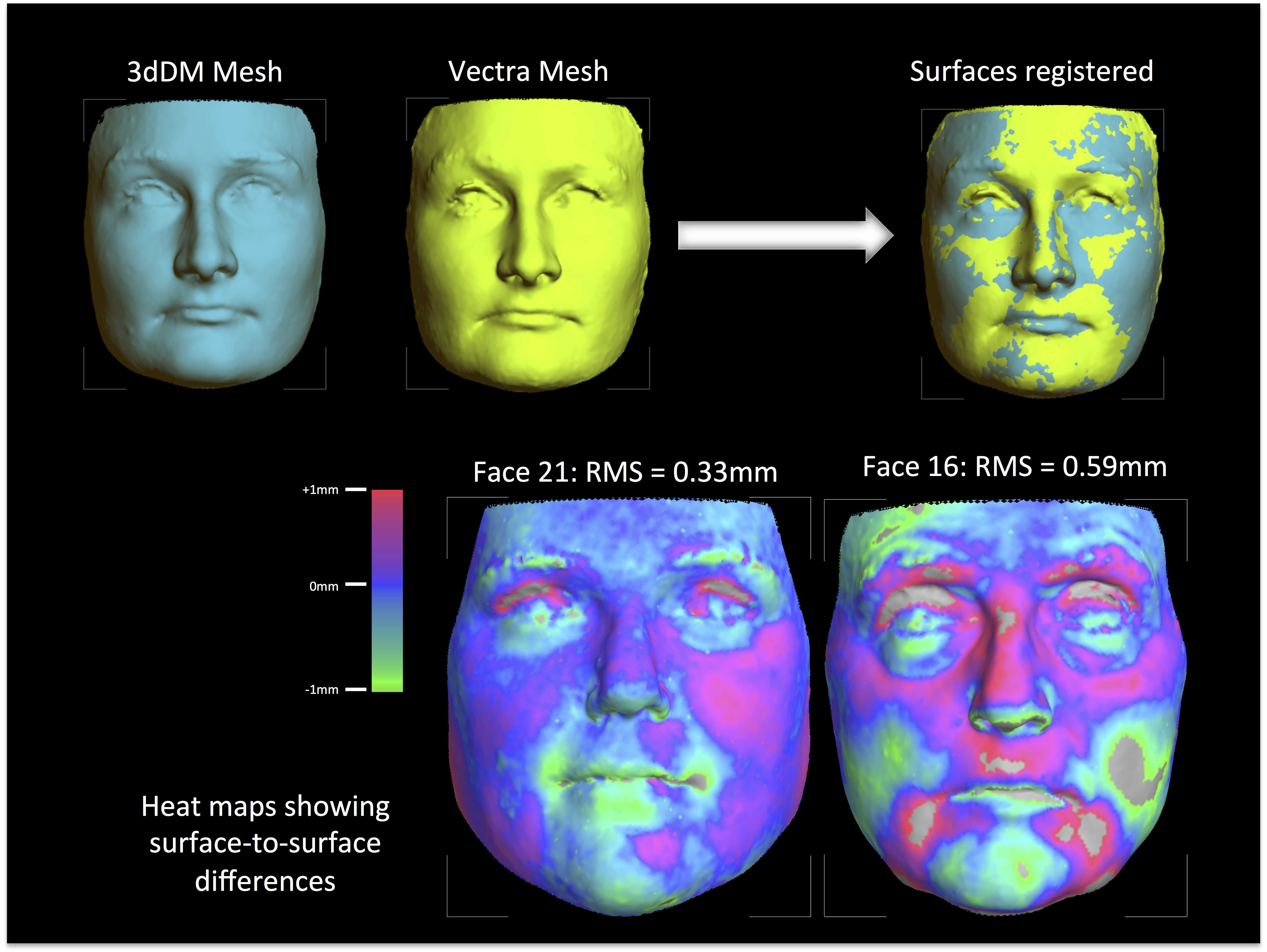
METHODS: 25 adult participants (age 21-62 years) were included in this IRB-approved study. Full frontal 3D facial images were obtained with both the 3dMDface system and the VECTRA H1 system under controlled conditions, carefully following manufacturer’s guidelines. The resulting surface models were trimmed to remove portions of the neck, shoulders and scalp hair. No surface smoothing or hole-filling routines were applied. Facial surfaces for each participant were registered to each other and heat maps were generated to visualize the magnitude and direction of the differences in geometry between the two surface models. This process involved an average 23,032 discrete point-to-point comparisons per face. Root mean square (RMS) error was calculated to assess the magnitude of the deviation in mm over the entire face.
RESULTS: The average RMS value of the 25 surface-to-surface comparisons was 0.43mm (range 0.33 - 0.59mm). In each case, the vast majority of the facial surface differences between H1 and 3dMD were within a +/- 1mm threshold. Areas exceeding +/- 1mm were generally limited to the upper eyelashes, eyebrows and corners of the mouth —regions containing hair and/or subject to facial microexpressions. There was no evidence of systemic directionally to the error, as the signed mean of the point-to-point differences was close to zero in each of the facial comparisons.
CONCLUSIONS: The validation of tools used to measure surgical outcomes is fundamental for high-quality practice and outcomes research. This study shows that 3D facial surface images acquired with the H1 system are accurate and comparable to the validated standard 3dMDFace system. This portability of the H1 comes at a cost, however, as the H1 requires several sequential captures of the face, whereas the 3dMDface system captures all angles simultaneously. The disadvantage of sequential capture is that subjects must maintain the same expression and position between images. At least in adults, this potential drawback did not result in major deviation in facial surface geometry.

Back to 2016 Annual Meeting
|
