|
|
|
|
|
Back to 2016 Annual Meeting
Evaluation of the structural integrity of the retina and optic nerve following whole eye transplantation using optical coherence tomography and manganese-enhanced magnetic resonance imaging
Chiaki Komatsu, M.D.1, Lin He, M.D.1, Yolandi van der Merwe, B.Eng.2, Maxine R. Miller, M.D.1, Katy A. Lucy, B.S.3, Huamin Tang, M.D.1, Ian Rosner, B.S.1, Yang Li, M.D.,Ph.D.1, Gadi Wollstein, M.D.3, Mario G. Solari, M.D.1, Joel S. Schuman, M.D.4, Kevin C. Chan, Ph.D.5, Kia M. Washington, M.D.6.
1Department of Plastic Surgery, UPMC, Pittsburgh, PA, USA, 2Department of Bioengineering, Swanson School of Engineering, University of Pittsburgh, Pittsburgh, PA, USA, 3UPMC Eye Center, Eye and Ear Institute, Ophthalmology and Visual Science Research Center, University of Pittsburgh School of Medicine, Pittsburgh, PA, USA, 4Department of Ophthalmology, New York University School of Medicine, New York, NY, USA, 5UPMC Eye Center, Eye and Ear Institute, Ophthalmology and Visual Science Research Center, Department of Bioengineering, Swanson School of Engineering, Pittsburgh, PA, USA, 6Department of Plastic Surgery, UPMC, VA Pittsburgh Healthcare System, Pittsburgh, PA, USA.
Background: The purpose of this study is to evaluate the viability and structural integrity of the visual system with in vivo optical coherence tomography (OCT) and manganese-enhanced magnetic resonance imaging (MEMRI) after whole eye transplantation (WET) in a rodent model.
Methods: Syngeneic transplants were performed in seven Lewis rats. The donor flap consisted of all ocular tissue anterior to the optic chiasm, a section of temporal bone and the skin of the eyelid and external ear. A similar region of the orbital contents was removed in the recipient. The recipient optic nerve was cut at the base of the globe. Anastomoses of carotid arteries and external jugular veins were performed, as well as optic nerve coaptation between donor and recipient. OCT was performed at one week after WET and MEMRI was performed at approximately three weeks after WET to qualitatively evaluate retinal structural integrity and anterograde Mn to evaluate retinal ganglion cell function.
Results: OCT revealed visualization of all layers of the retina in the transplanted eyes. Two of the rats had opacities of the cornea and thus there were inability to visualize their retina. Results from MEMRI of the naïve (untransplanted) contralateral eyes showed enhancement of the retina and intraorbital optic nerve, optic tract, lateral geniculate nucleus, and superior colliculus. MEMRI of the transplanted eyes revealed enhancement of the retina and intraorbital optic nerve up to a few millimeters behind the globe but not in the remainder of the visual pathway.
Conclusions: We have established a viable orthotopic whole eye transplant model in the rat. Qualitative analysis of OCT revealed that the structural integrity of the retina was relatively maintained after WET. MEMRI showed that anterograde Mn transport after WET was maintained in the donor eye to the site of transection at approximately three weeks after WET which may suggest some viability of retinal cells. Future studies will examine the exact physiological sources of manganese enhancement in the transplanted visual system, as well as approaches for neuroregeneration to regain eye-brain structural and functional connectivity in the WET model. 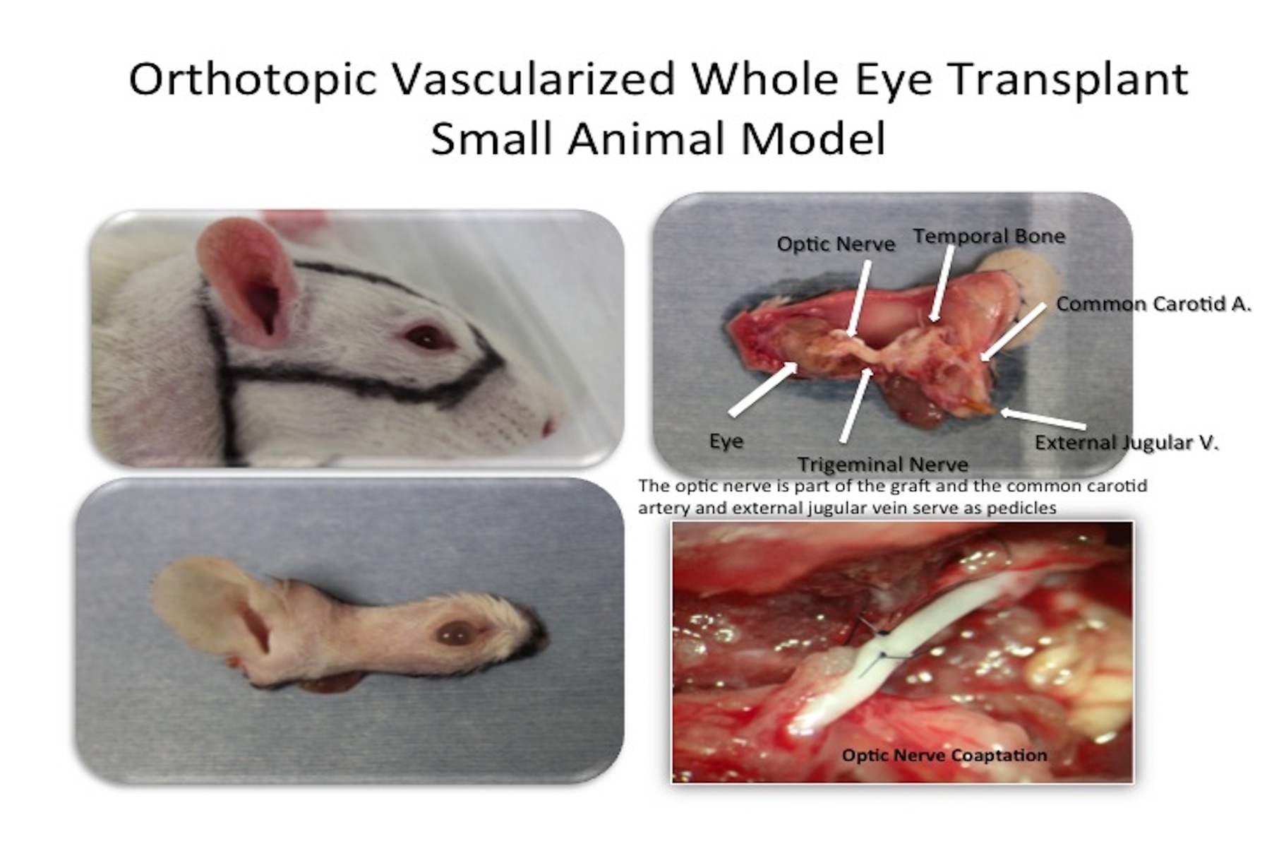 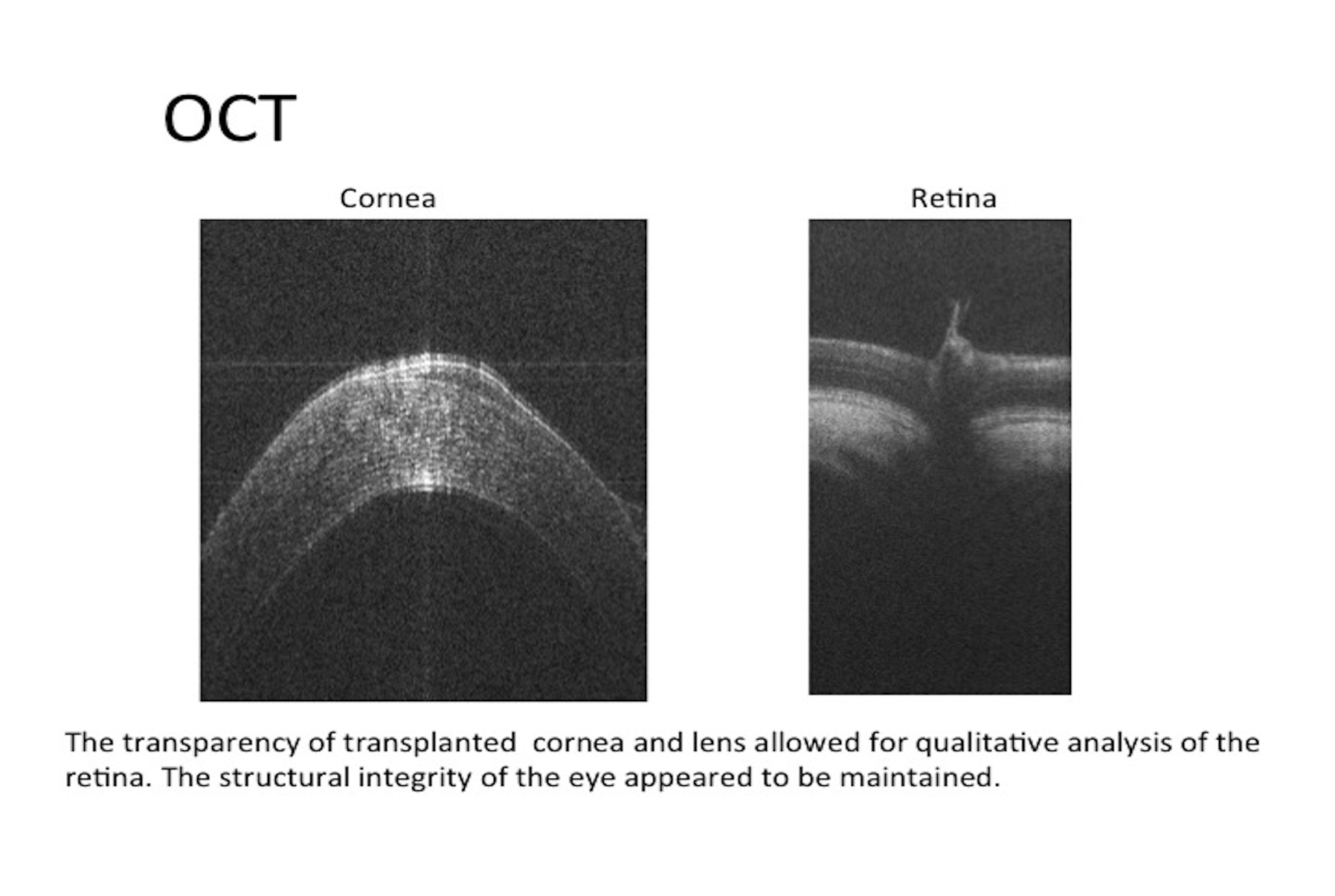 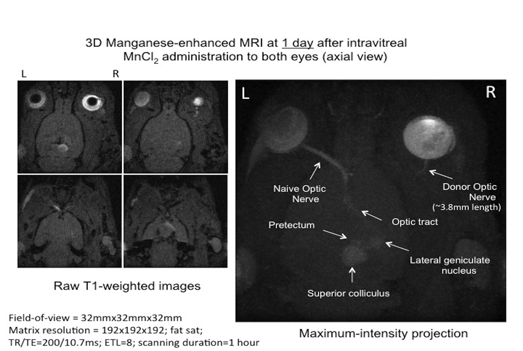
Back to 2016 Annual Meeting
|
|
