|
|
|
|
|
Back to 2016 Annual Meeting
Measuring Functional Recovery after Chronic Denervation of Peripheral Nerves: A Novel Rat Forelimb Model
Amy Quan, MPH1, Joseph Lopez, MD MBA1, Joshua Budihardjo, BS1, Sara Mermulla, MD1, Tariq A. Jawadi, MBBS2, Howard Wang, MD1, Ahmet Hoke, MD PhD1, Sami Tuffaha, MD1, W.P. Andrew Lee, MD1, Gerald Brandacher, MD1.
1Johns Hopkins Hospital, Baltimore, MD, USA, 2King Saud bin Abdulaziz University for Health Sciences, Riyadh, Saudi Arabia.
BACKGROUND: It is well understood that chronic denervation of upper and lower extremity motor nerves causes significant damage to peripheral nerves; Schwann cells (SC) undergo apoptosis, axonal basal lamina deteriorate, and target muscles atrophy. Thus, chronic denervation of peripheral nerves results in decreased nerve regeneration potential and negatively impacts clinical outcomes after extremity nerve trauma. Although the cellular and molecular effects of chronic denervation on peripheral nerve regeneration are well understood, previous studies have failed to correlate these results to behavioral functional results. To address this problem, we have developed a novel murine, upper extremity nerve injury model which optimizes measurement of functional recovery after chronic denervation injury.
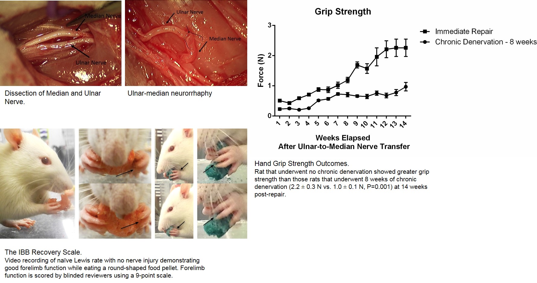
METHODS: We developed a forelimb chronic denervation model in which the median nerve was transected at the mid-humerus level and left in discontinuity for 8 weeks. After the period of chronic denervation was complete, the distal median nerve stump was co-apted to the proximal end of a freshly axotomized ulnar nerve. Group 1 rats underwent 8 weeks of chronic denervation (n= 8); Group 2 rats (positive control) did not undergo chronic denervation but instead underwent immediate nerve co-aptation of the median and ulnar nerves (n = 8); and Group 3 rats (naïve control) did not undergo any nerve procedure (n = 8). Functional recovery was tested weekly by measuring both grip strength using a force transducer and scoring the injured forelimb during feeding using a newly developed metric known as the IBB scale. Compound muscle action potentials (CMAPs) were also measured in the abductor pollicis brevis muscle of the injured limb at 8 and 12 weeks after nerve co-aptation/repair to provide a more objective metric to assess peripheral nerve regeneration. Animals were sacrificed at 14 weeks after ulnar-median neurorrhaphy for assessment of axonal regeneration and degree of muscle atrophy.
RESULTS: Fourteen weeks after ulnar-median neurorrhaphy, Group 2 rats demonstrated significant functional recovery, regaining 63% of its baseline grip strength, 78% of its baseline feeding score, and 46% of its baseline CMAPs. At 14 weeks, Group 1 rats demonstrated less functional recovery: regaining 25% of its baseline grip strength, 67% of its baseline feeding score, and 15% of its baseline CMAPs. Group 2 animals demonstrated greater grip strength than the Group 1 animals (2.2 ± 0.3 N vs. 1.0 ± 0.1 N, P=0.001), improved feeding (score of 7.3 ± 0.05 vs. 6.0 ± 0.04, P<0.0001), and greater CMAPs (1.7 ± 0.07 millivolts vs. 0.5 ± 0.04 millivolts, P=0.0001). Furthermore, forelimb flexor muscle weights were significantly different between Group 2 and 1 (0.82g vs 0.53g; p < 0.00001).
CONCLUSIONS:This novel forelimb chronic denervation model provides the first translatable animal model to assess functional recovery after peripheral nerve chronic denervation injury. This model provides a reliable model to assess future therapeutics aimed at augmenting extremity function following chronic denervation injury of peripheral nerves.
Back to 2016 Annual Meeting
|
|
