
Analysis of Follicle-Stimulating Hormone Receptor Expression in Infantile Hemangioma
Reid A. Maclellan, M.D., M.M.Sc, Javier A. Couto, MD, Fu Xi, MD, Lan Huang, PhD, Joyce Bischoff, PhD, Arin K. Greene, MD, MMSc.
Children's Hospital Boston / Harvard Medical School, Boston, MA, USA.
Background: The life-cycle of infantile hemangioma and follicle-stimulating hormone (FSH) secretion are identical. We have shown that infantile hemangioma expresses the receptor for follicle-stimulating hormone (FSHR). The purpose of this study was to identify which cell type(s) in infantile hemangioma contain FSHR.
Methods: Human infantile hemangioma sections and cells were subjected to immunofluorescence for FSHR. Tissues were co-stained with DAPI and either anti-PDGFR-β or anti-CD31 antibodies to identify nuclei, pericytes, and endothelial cells, respectively. Specimens also were fractionated by fluorescence-activated cell sorting (FACS) into hematopoietic, endothelial, perivascular, and mesenchymal stem cells and were tested for the presence of FSHR by using FSHR antibody. Control specimens consisted of sertoli cells (positive) and normal skin/subcutis (negative).
Results: FSHR was expressed in the endothelial, perivascular, and stem cells of infantile hemangioma by immunofluorescence and FACS. Receptor expression by FACS was greatest in stem cells (37.2%), compared to pericytes (13.5%) or endothelial cells (8.4%) (Figure).
Conclusions: Endothelial cells, pericytes, and stem cells in infantile hemangioma express FSHR. A precursor cell giving rise to endothelial cells and pericytes expressing FSHR might contribute to the pathogenesis of infantile hemangioma.
Figure: Fluorescence-activated cell sorting demonstrates (A) 56.9% and (B) 23.6% follicle-stimulating hormone receptor expression in stem cells from two individual proliferating infantile hemangioma specimens. 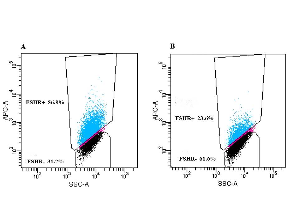
Back to 2017 Program


