
A More Clinically Relevant Rodent Model of Peri-Prosthetic Capsular Contracture
Matthew A. Wright, BA, Arash Samadi, BS, Daniel O. Lara, BS, Samantha Shelffo-McGrath, Alexandra J. Lin, BA, Sarah J. Karinja, MD, Jason A. Spector, MD.
Laboratory of Bioregenerative Medicine and Surgery, Weill Cornell Medical College, Department of Surgery, Division of Plastic Surgery, Cornell University, New York, NY, USA.
Introduction: Capsular contracture is the most common complication of breast implantation. Multiple animal models have been utilized to study this complication, including mouse, rat, rabbit, and pig models. In nearly all of these studies, the devices are placed subcutaneously. While making for a simple implantation procedure, the subcutaneous model is not the most accurate recapitulation of clinical practice. Furthermore, subcutaneous placement results in less reliable histologic analysis and capsule thickness measurements given that there is already abundant connective tissue in this plane at baseline. We have developed a rat model of breast implant placement - in which smooth round silicone implants are placed under the latissimus dorsi muscle - that not only more closely models usual clinical practice but allows for more accurate assessment of peri-prosthetic capsule thickness.
Methods: Four female Sprague Dawley rats were split into two groups and implanted with either subcutaneous or submuscular 2cc smooth silicone implants (Mentor). Subcutaneous implants were placed in pockets dissected bilaterally on the dorsum. For submuscular implantation, the lateral edge of the latissimus dorsi muscle was identified, an implant was placed under the latissimus, and the lateral edge of the latissimus was tacked back down to the chest wall (Figure 1, a-b). A rat from each group was sacrificed either immediately or at one month, and capsular histology was examined.
Results: Analysis of subcutaneously placed implants explanted immediately postoperatively demonstrated a significant layer of connective tissue between the implant and the overlying panniculus carnosus muscle (i.e. where the capsule will form). This layer was as thick as 500 μm (the capsule itself may be significantly thinner) at some points and nearly identical in histologic appearance to capsule. Analysis of submuscularly placed implants explanted immediately postoperatively demonstrated no connective tissue between the implant and the overlying latissimus dorsi muscle. At one month, capsule had clearly formed in the rats from both groups, but true capsular tissue was much more readily identified in the submuscular versus the subcutaneous plane (Figure 1, c-f).
Conclusions: Submuscular placement under the latissimus dorsi makes for a more clinically relevant and histologically accurate model of capsular contracture as it obviates the often indistinct border between native subcutaneous fascia and peri-prosthetic capsule seen with subcutaneous placement that can lead to inaccurate assessments of capsule thickness. We believe this method represents the gold standard for the study of breast implantation using a rodent model. 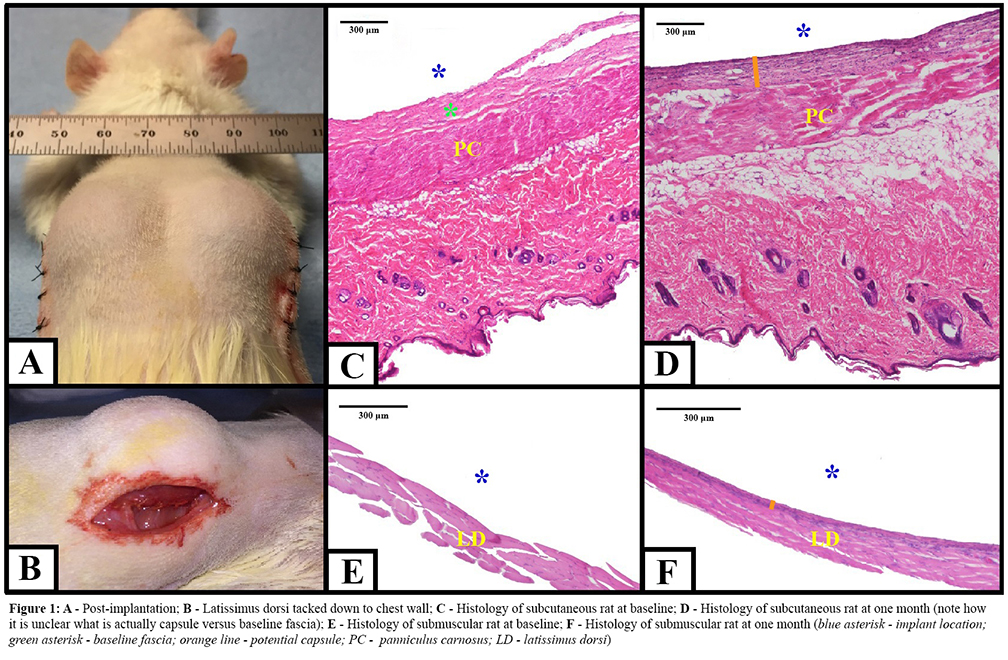
Back to 2018 Posters


