
Indocyanine green microangiography in the cadaveric model: a technique for microsurgical simulation
Chrisovalantis Lakhiani, MD, Tara M. Chadab, MS, David Janhofer, BS, Karen K. Evans, MD, David H. Song, MD, MBA.
Georgetown University Hospital, Washington, DC, USA.
Background
Realistic microsurgical simulation models can help surgical residents practice technically demanding skills in safe environments. Prior studies have used indocyanine green (ICG) microangiography to mimic physiologic tissue perfusion, in the setting of an artificially perfused cadaveric system, for simulation of microvascular procedures.1,2 However, ICG microangiography has not been attempted for the in-situ assessment of raised flaps in the cadaveric model. Here we attempt to show the feasibility and reliability of ICG microangiography as a method of flap assessment in the cadaveric model.
Methods
Three fresh frozen cadavers were obtained from our institution’s willed body program. After flap dissection was performed, the afferent vessel was cannulated with an 18 or 22-gauage IV catheter. A one-liter solution of lactated ringer’s solution on a pressure bag, or via manual compression, was hung and connected to the IV tubing. The ICG was administered as a secondary infusion into the flap and allowed to drain via the efferent vessel into a collecting basin. The SPY Elite imaging system was used to capture and record the fluoroscopic image.
Results
Two mid-level plastic surgery residents participated in raising cadaveric flaps. Flaps raised included 5 anterolateral thigh, 5 superficial circumflex iliac artery perforator, and 5 radial forearm flaps. Perfusion was then assessed using indocyanine green angiography. Of 15 flaps, 13 were raised successfully and 2 were not owing to technical misadventure, which was identified by ICG leakage through transected or avulsed vessels. This provided opportunity for teaching and refinement in technique.
Conclusion
ICG microangiography is a feasible and reliable method of assessing flap perfusion in the cadaveric model. Consequently, it can be a valuable tool for practicing microsurgical techniques outside of the operating room.
References
1. Carey JN, Rommer E, Sheckter C, Minneti M, Talving P, Wong AK et al. Simulation of plastic surgery and microvascular procedures using perfused fresh human cadavers. J Plast Reconst Aesthet Surg. 2014;67(2):e42-8.
2. Carey JN, Minneti M, Leland HA, Demetriades D, Talving P. Perfused fresh cadavers: method for application to surgical simulation. Am J Surg. 2015;210(1):179-87.
Figure 1. Superficial Circumflex Iliac Artery Perforator (SCIP) Flap 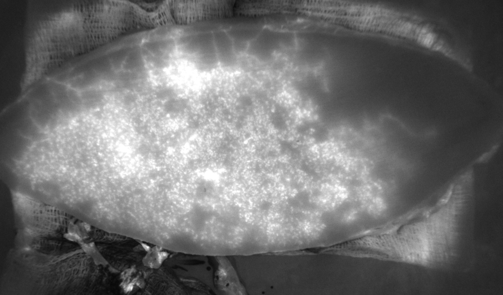
Back to 2018 Posters


