
Timing of Lymphatic Imaging Using Indocyanine Green (ICG) Lymphangiography in Normal Upper Extremities
Elizabeth O. Kenworthy, MD, Itay Wiser, MD, PhD, Babak Mehrara, MD, Joe Dayan, MD.
Memorial Sloan Kettering, New York, NY, USA.
Introduction:
Indocyanine green (ICG) lymphangiography has become routinely used in evaluating patients for lymphatic surgery yet there is currently minimal published literature on the normal anatomy of the lymphatic system using ICG. While definitions of abnormal findings on ICG have been elucidated, a description of the normal anatomy is important in order to better understand, diagnose, and effectively treat lymphedema. The purpose of this study was to evaluate ICG findings in the normal upper limb.
Methods
ICG lymphangiography of the upper limb was evaluated in healthy consecutive patients. Dynamics of lymphatic uptake were measured at the wrist, antecubital fossa, upper arm, axilla and supraclavicular lymph nodes by taking ICG images at 5, 30 and 60 minutes following injection. Paired T-test was used to compare lymphatic vessel count between various injection sites. Sign rank test was used to compare time point measurements.
Results
Initial data on 25 normal upper extremity ICG images in 24 patients were reviewed. Sixteen patients (64%) showed uptake in the axilla after five minutes, while all upper extremities showed uptake into the axilla at 30 minutes after injection. Four upper limbs (20%) had uptake in the supraclavicular lymph nodes at 60 minutes after injection. Time after injection was found to be significant in observing more lymphatics in the wrist, forearm, and upper arm in the 5 minute to 30 minute images (p=0.003, p=0.000, p=0.000 for wrist, antecubital and upper arm respectively)
and 5 minute to 60 minute images (p=0.000, p=0.000, p=0.000 for wrist, antecubital and upper arm respectively). There was no statistical difference in the upper arm between 30 and 60 minutes (p=0.500).
Conclusion
ICG lymphangiography provides a detailed view of the lymphatic system, allowing for direct assessment in the clinic. While more lymphatics are visualized at 30 and 60 minutes after injection, the clinical significance of this remains unknown. The work presented in this paper describes normal anatomy by which abnormal findings can be compared. This may serve as a starting point for further work in using ICG as a validated diagnostic and assessment tool. 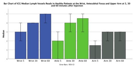
Back to 2018 Posters


