
Silicone Implant Shells Drive Proliferation of Patient-Derived BIA-ALCL Cells in Engineered Biomimetic Patient-Derived Breast Tissue
Ishani D. Premaratne, BA1, Matthew A. Wright, BA2, Runlei Zhao, MD1, Daniel O. Lara, BA1, Arash Samadi, BA1, Cosima S. Boettner3, Giorgio Inghirami, MD1, Kristy A. Brown, PhD1, Jason A. Spector, MD4.
1Weill Cornell Medicine, New York, NY, USA, 2Columbia University Vagelos College of Physicians and Surgeons, New York, NY, USA, 3Tufts University, Boston, MA, USA, 4Weill Cornell Medicine, Department of Surgery, New York, NY, USA.
Background: Breast implant-associated anaplastic large cell lymphoma (BIA-ALCL), a rare but potentially deadly complication of device-based breast reconstruction, has an estimated incidence of 1 in every 3817 cases of textured device implantation. Present hypotheses of BIA-ALCL pathogenesis are difficult to test due to the inadequacy of in vivo and in vitro models. This study utilizes a high-fidelity ex vivo biomimetic, three-dimensional model to study the effects of implant shells on patient-derived BIA-ALCL cells within the breast microenvironment.
Methods: Patient-derived breast tissue was processed for its component adipocytes, organoids, and stromal vascular fraction. These were suspended within 50 µl of 0.3% type I collagen matrix with patient-derived BIA-ALCL cells at a density of 200,000 cells/mL before being plated into 6mm wells. As a control, BIA-ALCL cells were suspended within type I collagen at the same seeding density and volume without breast components. Before plating, wells were lined with either textured, smooth, or no implant shells. These were 1cm by 2cm pieces of implant shell dissected from the whole implant, cleaned of underlying silicone gel, and autoclaved before being placed into the wells with the superficial aspect of the shell facing inward. Eight wells were plated per implant shell type: four biomimetic platform wells and four collagen-only controls. All groups started at an equal density of 1000 ALCL cells per confocal snapshot. Wells were imaged immediately and every other day using confocal microscopy before being processed using Imaris™ to analyze cell proliferation over time.
Results: BIA-ALCL cell proliferation was significantly more robust in the biomimetic platform relative to the collagen-only groups regardless of implant shell type (p-value less than 0.01). BIA-ALCL cells in textured and smooth shell biomimetic groups grew nearly 30% faster than those within biomimetic wells lacking implant shell, with statistical differences as early as day two. After ten days in culture, mean cell counts in the textured and smooth shell biomimetic groups were 4021±999 and 4281±633, respectively, compared with 2399±355 in the biomimetic group lacking implant shell (p=0.015). There was no statistical difference in BIA-ALCL cell proliferation between the textured and smooth biomimetic groups or among any of the collagen-alone groups.
Conclusions: Using a tissue-engineered three-dimensional ex vivo model of BIA-ALCL, we have demonstrated that BIA-ALCL cells thrive within the biomimetic platform when compared to collagen alone and that incorporation of smooth and textured implant shell leads to significantly increased BIA-ALCL cell proliferation compared with no implant shell. 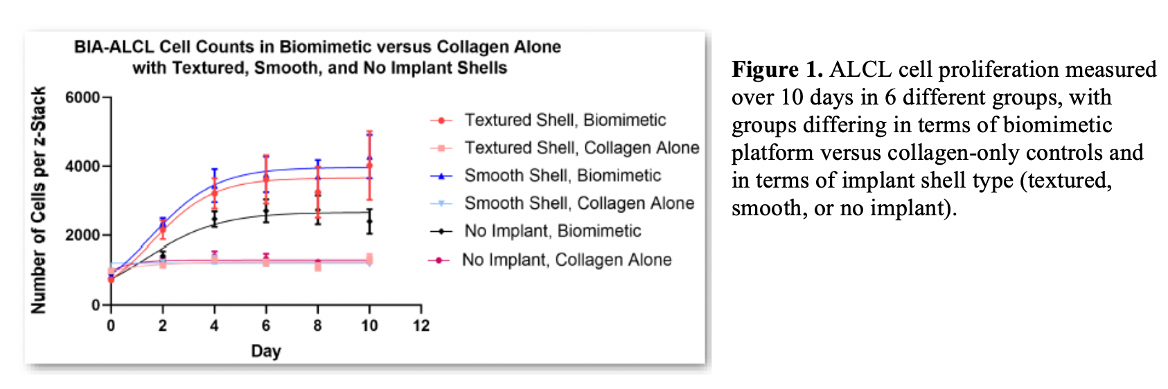
Back to 2019 Abstracts