
How Does the Bandeau Grow: Quantifying Post-Operative Changes in the Bandeau Over Time after Fronto-orbital Advancement
Lucas A. Dvoracek1, Aaron Foglio1, Sean Herman, MD1, Eva Roy1, Scott P. Bartlett, MD2, Joseph E. Losee, MD1, Jesse A. Taylor, MD2, Jesse A. Goldstein, MD1.
1University of Pittsburgh, Pittsburgh, PA, USA, 2University of Pennsylvania, Philadelphia, PA, USA.
BACKGROUND: Temporal hollowing is a late sequelae of fronto-orbital advancement (FOA) surgeries which contributes to aesthetic deformity and patient dissatisfaction. Bony manipulation including devascularization and under-correction may contribute to temporal hollowing. We sought to objectively assess how such bony morphology changes over time. METHODS: A multi-center, retrospective study identified craniosynostosis (CS) patients treated with FOA between 2008-2018 with both early and late (>12 mos) post-operative head CT scans. Scans were reconstructed, oriented, and manually segmented into surgical fragments that delineated the osteotomies of interest for a given patient. Symmetric and asymmetric bandeaus were subdivided into hemibandeaus for comparison. Hemibandeaus were evaluated for 32 coordinate points and 35 discreet metrics, and measurements compared between timepoints for a given Hemibandeau. RESULTS: Twenty patients matched inclusion criteria (12 female:8 male). CS subtypes included metopic (7), unilateral coronal (6 right, 3 left), and other (4). Median age at surgery and time to follow-up scan was 1 and 2.1 years respectively. 36 hemibandeaus were identfied Average cranial width and length growth and was 1.2%/year and 3.7%/year respectively. The bandeau AP length increased 7.2%/year and while average bandeau posterior width increased 2.0%/year, anterior width decreased by 0.4%/year (not significant), leading to a relative anterior transverse deficiency. The average initial bandeau orbital width was 3.6mm wider than the midface orbital width and decreased by 1.2mm (~36% loss of overcorrection).
CONCLUSIONS: The long-term shape and position of the bandeau determines surgical success of FOA. Although the bandeau continues to grow comparably to native skull in some dimensions after surgery, quantifiable deficiency in growth occurs in the anterior temporal and zygomatico-frontal regions. This is the first comparative demonstration of the bony contribution toward temporal hollowing in early and late post-operative patients. 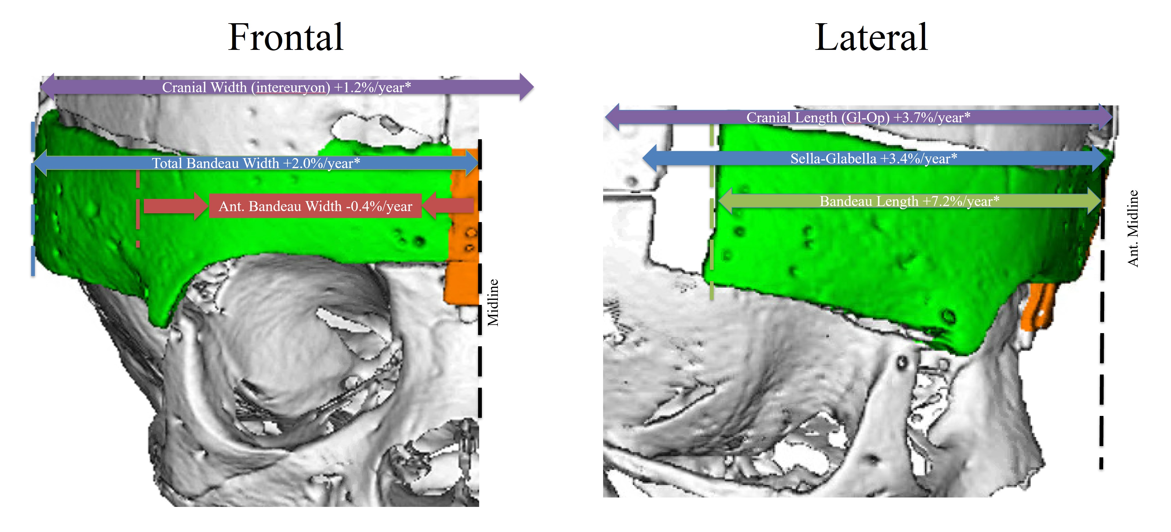
Back to 2019 Abstracts