
Successful Creation of Lymphedema in a Rat Model
Jeffrey A. Ascherman, MD, Amro A. Harb, BA, Celine F. Nicolas, BS, John J. Corvi, BS, Maxwell A. Levi, BS, YuanDian Zheng, BS, Yelena Akelina, DVM, MS, Eileen P. Connolly, MD, PhD.
Columbia University Irving Medical Center, New York, NY, USA.
Background: Chronic lymphedema is a condition of lymph accumulation due to impaired lymphatic drainage. An increasingly popular technique to treat lymphedema is the vascularized lymph node transfer (VLNT), but there has been a lack of animal studies to evaluate this treatment, partially due to the difficulty of creating a rat model of lymphedema. The purpose of this experiment was thus to create a rat lower limb lymphedema model.
Methods: Inguinal and popliteal lymphadenectomies were performed in 5 Sprague-Dawley rats. The inguinal and popliteal nodes were identified using isosulfan blue dye and excised with their surrounding adipose tissue. A 5-10 mm open integumentary defect was created around the circumference of the proximal thigh, and skin edges were sutured to the underlying muscle to disrupt dermal and subcutaneous lymphatic flow. After 3 weeks, rats were administered a single, unfractionated 22.7 Gy dose of radiation at a rate of 1 Gy/min in a field size of 4x4 cm. Monthly bioimpedance and ankle circumference measurements were obtained. At approximately 4 months post-radiation, indocyanine green (ICG) dye was injected intradermally in the feet to visualize lower extremity lymphatic drainage.
Results: Three of the 5 rats lived at least 4 months following the procedure. Two of these 3 had average bioimpedance ratios less than 0.9 (0.774 and 0.823), consistent with lower extremity lymphedema. In one of these rats, ICG lymphangiography revealed diffuse lymphatic drainage up to the proximal thigh, but not beyond the thigh, consistent with lymphedema. This animal’s contralateral (non-operated) leg had normal lymphatic drainage across the thigh into the inguinal node (Figure 1). Ankle circumference measurements of the operated and non-operated legs at 4 months postoperatively were 40 and 31 mm respectively. In the other rat with a bioimpedance measurement consistent with lymphedema, ICG lymphangiography also showed abnormal lymphatic drainage in the operated leg, and ankle circumference measurements of the operated and non-operated legs were 36 and 32 mm, respectively.
Conclusions: In our study, inguinal and popliteal lymphadenectomy, combined with disruption of skin and subcutaneous lymphatic drainage in the proximal thigh, and unfractionated radiotherapy, successfully created lymphedema in 2 of 3 rats at 4 months postoperatively. We believe this rat lymphedema model will be very helpful for future lymphedema studies, including of VLNT as a treatment for lymphedema.
Figure 1: ICG Image 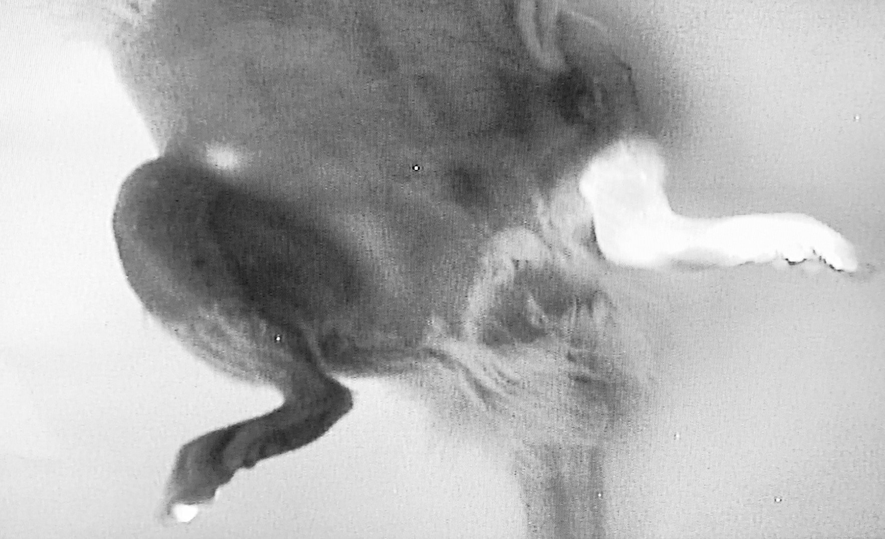
Diffuse dermal backflow is visualized in the operated left leg; linear drainage into the inguinal node is visualized in the non-operated right leg.
Back to 2019 Abstracts