
FlapMappreoperative surgical planning system for deep inferior epigastric perforator flap breast reconstruction
Farah Sayegh, MD1, Taylor Ibelli, BS, MS2, Alexander Kagen, MD1, Peter W. Henderson, MD1.
1Icahn School of Medicine at Mount Sinai, New York, NY, USA, 2Sackler School of Medicine at Tel Aviv University, Tel Aviv, Israel.
BACKGROUND: Preoperative imaging of the lower abdominal vascular and structural anatomy has been shown to reduce operative time and enhance surgical outcomes in deep inferior epigastric perforator (DIEP) flap breast reconstruction. Merely having raw, unanalyzed imaging data, however, does not constitute a preoperative surgical plan. Therefore, the goal of this project was to develop a means of creating a well-defined DIEP flap surgical plan utilizing data from preoperative tomographic imaging.
METHODS: Raw images from tomographic imaging series generated for preoperative vascular mapping were analyzed, and extracted data involved rectus abdominis muscle size, location, and inscription anatomy, perforator and pedicle location and three-dimensional course, and superficial venous drainage patterns. Relevant data points were identified, measured, translated to a newly developed visual language pattern, and plotted on a Cartesian coordinate grid.
RESULTS: The resulting clinical device is the FlapMap, a novel DIEP flap surgical planning system that condenses into a single image the large amount of clinically relevant data contained within a stack of preoperative tomographic images. The creation of this customized plan requires eight straightforward steps. The relative depths of the pedicle segments are color-coded as green (submuscular), red (intramuscular), and purple (subfascial/supramuscular); and the superficial venous system is denoted in blue. A sample template can be downloaded at http://bit.ly/FlapMaptemplate.
CONCLUSIONS: We present for the first time the FlapMap system for preoperative planning for DIEP flap breast reconstruction. By analyzing multiple tomographic images in a standardized and methodical manner, clinically relevant information can be extracted and synthesized in order to create a single, standardized image that contains actionable information that can lead to the development of a preoperative surgical plan. Such planning has the potential to improve both systems measures (such as operative time) and patient outcomes (such as complications and microsurgical patency rates). This system has many potential future developments, including process automation through machine learning, and intraoperative visualization and navigation capabilities. 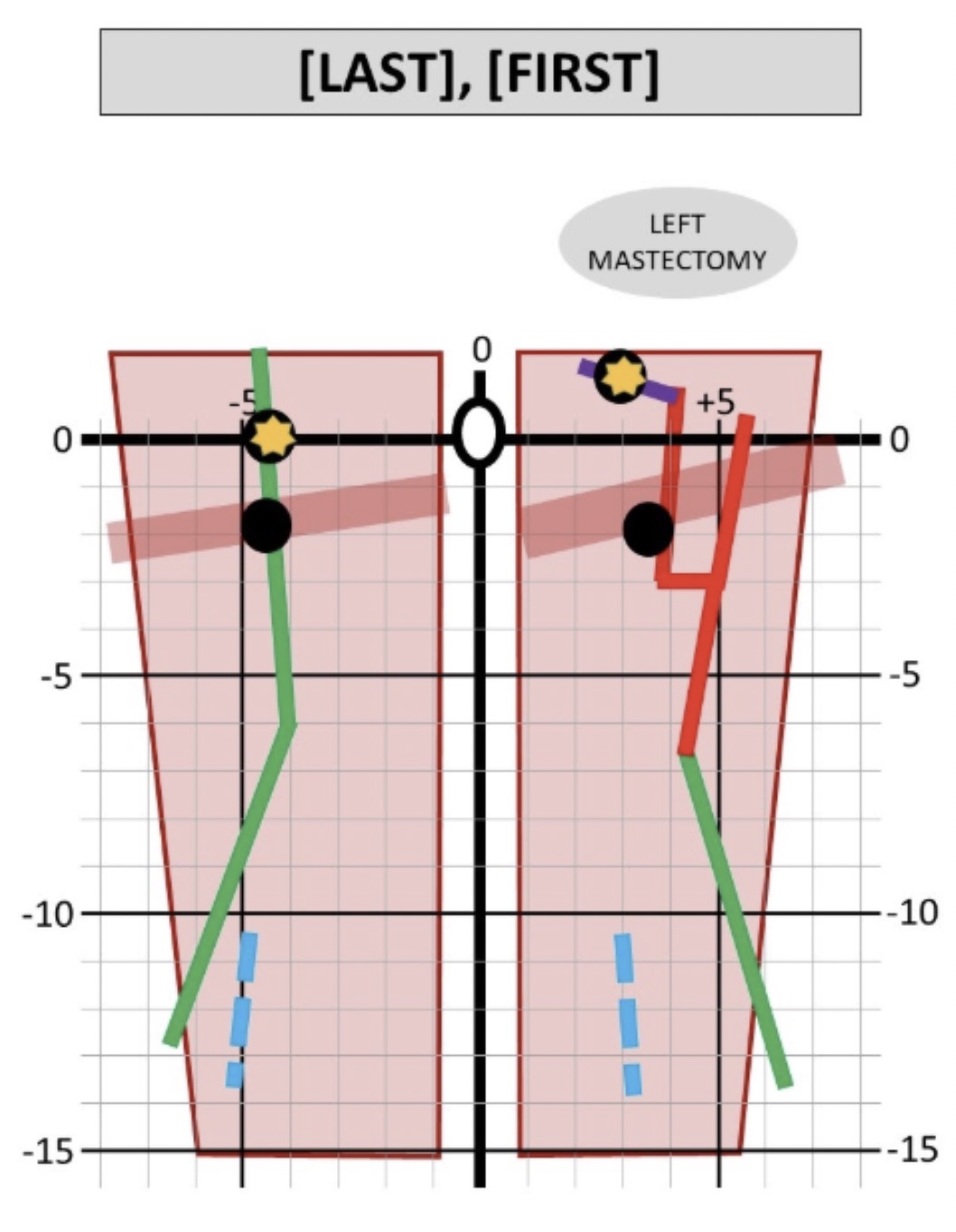
Back to 2020 Abstracts