
Evolution of Anomaly-Specific Techniques in Infant Ear Molding: A Ten-Year Retrospective Study
Karina Charipova, BS1, Ashley Rogers, MD2, Manas Nigam, MD2, Vikas S. Kotha, BS3, Christina Barra, NP2, Stephen B. Baker, MD, DDS2.
1Georgetown University School of Medicine, Washington, DC, USA, 2Department of Plastic Surgery, MedStar Georgetown University Hospital, Washington, DC, USA, 3George Washington University School of Medicine and Health Sciences, Washington, DC, USA.
BACKGROUND: Congenital ear anomalies occur in at least one-third of the population, and less than one-third of cases self-correct.1 Ear molding is an alternative to surgery that spares operative morbidity and allows for significantly earlier intervention. This study describes prosthetic techniques developed by the senior author to adapt standard correction systems to each specific type of ear anomaly.
METHODS: The authors conducted a retrospective, institutional review board-approved study of 246 patients who underwent ear molding by a single surgeon. Each case was reviewed to develop step-wise customization protocols for existing EarWellTM and InfantEarTM systems.
RESULTS: This review included 385 ears in 246 consecutive infants who underwent ear molding between January 2010 and December 2019. Patient age at presentation ranged from less than 1 week to 22 weeks. Presenting ear anomalies were subclassified into Stahl’s ear (3.6%), lidding/lop (9.3%), helical rim (28.5%), prominent (10.6%), cupping (2.8%), conchal crus (3.3%), and mixed (37.4%). Anomaly subclass could not be obtained for 11 patients (4.5%). At the end of this ten-year experience, the senior author was implementing modifications in over 30% of patients (Figure 1). Recommended modifications to existing ear correction systems are anomaly-specific: depression of the third crus with a cotton tip applicator (CTA) set with dental impression material or silicone gel (Stahl’s ear); molding of scaphal form with soldering wire secured with dermal glue, dental impression material, or silicone gel (helical rim/lop); posterior depression of the conchal bowl with a CTA set in silicone gel (prominent); retraction of the earlobe with dermal glue applied to the posterior edge of the lobe (isolated projecting earlobe); creation of a custom stent using dental impression material with implementation of a long-term retainer made of dental putty following completion of treatment (cupping/lidding/lop/conchal crus); and weekly, dynamic repositioning of retractors under the superior cartilage (cryptotia). Of surveyed parents, 92% expressed satisfaction with outcomes of treatment.
CONCLUSIONS: Presentation of ear anomalies is heterogenous. This ten-year experience demonstrates that the approach to ear molding should be dynamic and customized, using techniques beyond those listed in system manuals to complement each ear and to improve outcomes.
REFERENCES
1. Chan S, Lim G, Por Y, et al. Efficacy of ear molding in infants using EarWell infant correction system and factors affecting outcome. Plast Reconstr Surg. 2019;144(4):648-658.
Figure 1. Use of a cotton tip applicator in combination with an EarWell to correct a helical rim-scaphal deformity. 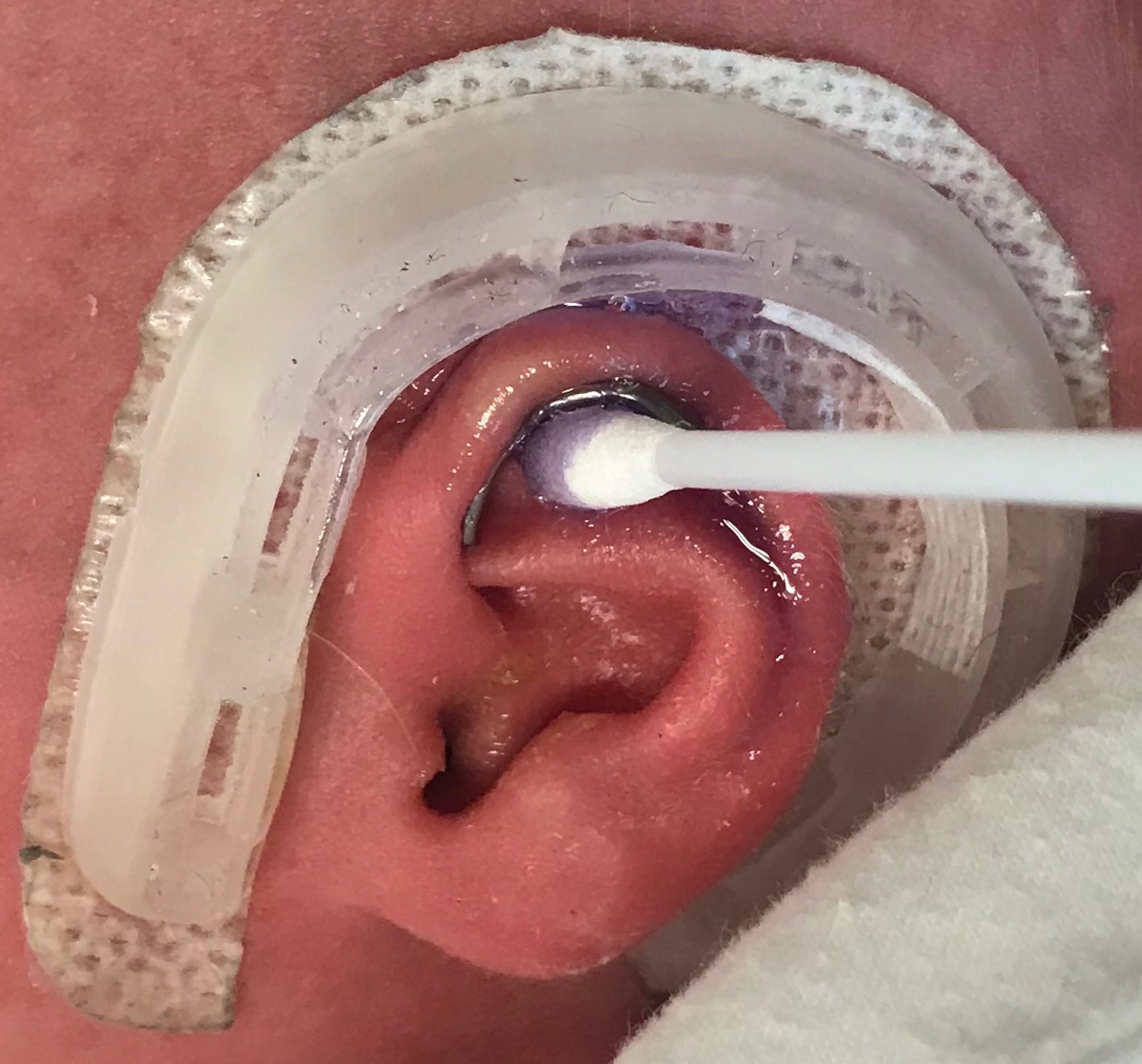
Back to 2020 Abstracts