
Anterior Ear Reconstruction with the Posterior Pull-Through Flap
John E. Gatti, MD, FACS, Robert B. Sollitto, MD, FASDS
Voorhees Surgery Center, Voorhees, NJ
BACKGROUND: Defects of the central ear with exposed cartilage after skin cancer removal remains a common problem for the reconstructive surgeon. The experience with a one-stage, post-auricular, skin-island flap passed through the cartilage to reconstruct ear defects is reported.
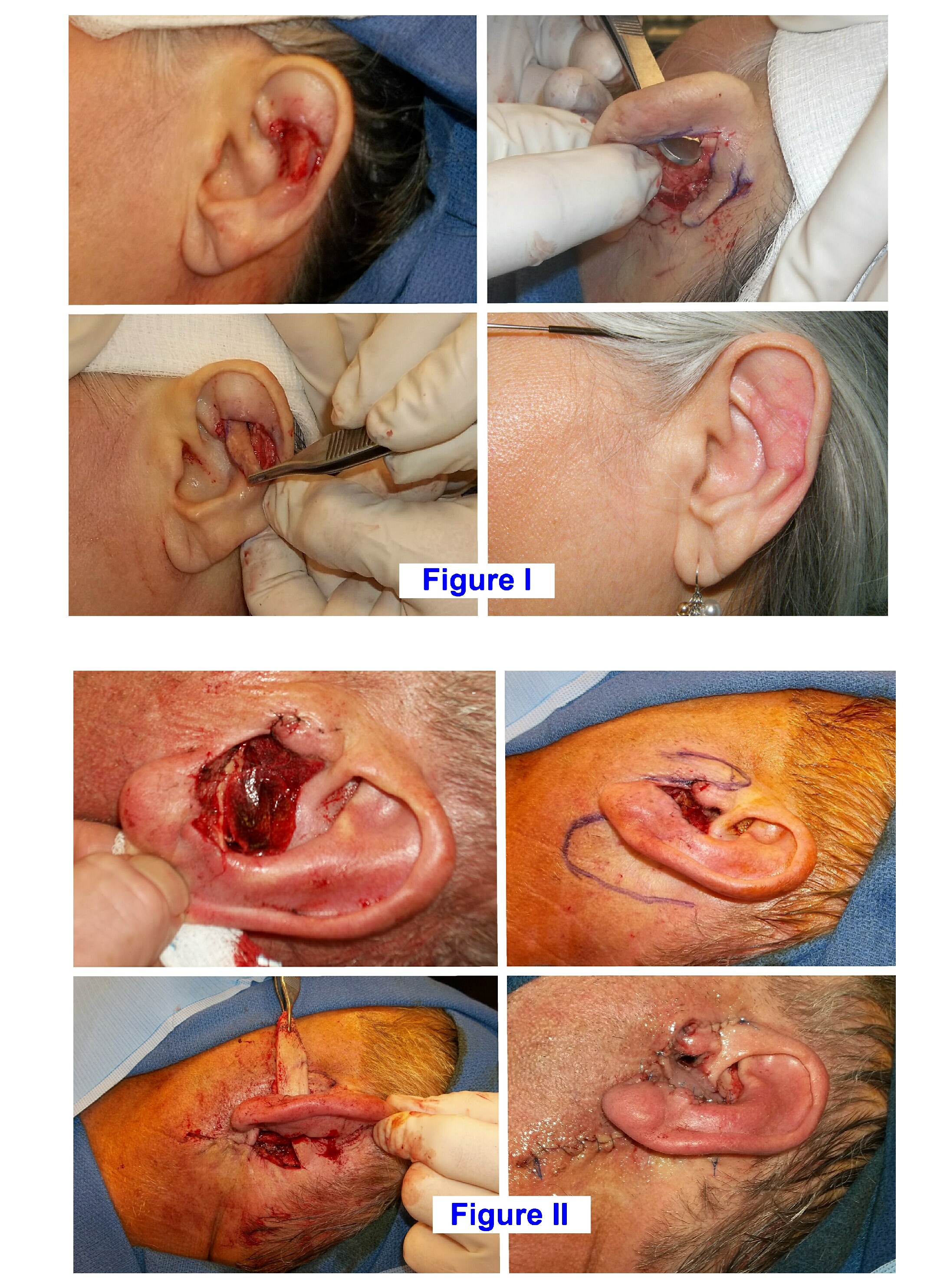
METHODS: Patients with an intact ear helix and an anterior full-thickness defect were reconstructed with a skin-island flap based on posterior subcutaneous tissue deep to the flap base. The flap was passed through a generous aperture created in the ear cartilage to ensure adequate perfusion and avoid congestion. The epithelium was incised superficially to create a distal skin island. The edge was lifted slightly from the subcu tissue and the flap inset to fill the defect (Figure I). All epithelium was secured exteriorly and not buried. The flap was secured at its periphery with absorbable chromic sutures. The post-auricular harvest site was closed primarily with chromic sutures. Minimal dressings and no external stentings were utilized with the reconstructions. Small chromic sutures were occasionally used across the flap to eliminate space beneath the flap. Antibiotics were administered pre-operatively and for 5 days post-operatively.
RESULTS: Twenty-two patients (6 women, 16 men) with anterior ear defects underwent single-stage reconstruction with posterior pull-through flaps over a 9-year period. The defects reconstructed measured in length from 2 cm to up to 4.5 cm. Five patients required a second flap from pre-auricular skin to help close ear canal defects (Figure II). All flaps adequately closed the defects with this one stage reconstruction, added structural support, and prevented ear distortion. Occasionally, venous congestion was observed in the distal flap but it was self-limiting and full thickness necrosis did not occur. The chromic sutures dissolved within a few weeks; facilitated by patient application of antibiotic ointment to the areas and light cleansing. No epithelial cysts developed. Ear position remained normal and ear projection was not observed.
DISCUSSION: Skin cancer extirpation of the anterior ear often exposes cartilage and leaves structural defects within the central ear. A one-stage post-auricular flap can reliably reconstruct defects and add support to the ear. Large defects may be closed without distorting shape or position of the ear. Care is needed to provide a generous aperture through the ear cartilage for passage of the flap base. Healing proceeds predictably and minimal complications are associated with this posteriorly based, pass-through, skin-island flap in ear reconstruction.
Back to 2022 Abstracts