
Taking Micrognathia On The Chin: Fabrication Of Customized, 3D-printed Genial Implants From A Novel Means Of Three-Dimensional Virtual Surgical Planning
Nicholas A. Vernice, A.B.1, Carly Askinas, B.S.1, Sabrina Shih, B.A.1, Xue Dong, M.D, Ph.D.1, Gillian O'Connell, A.B.,1 Ryan J. Bender, B.S.,1 James Shin, M.D.2, Jason A. Spector, M.D.1,3
1Division of Plastic and Reconstructive Surgery, Weill Cornell Medicine, New York, NY USA2Department of Radiology, Weill Cornell Medicine, New York, NY USA3Department of Biomedical Engineering, Weill Cornell Medicine, New York, NY USA
Background: Surgical alterations of the chin are increasingly common, with indications varying from the correction of congenital hypoplasia to cosmetic enhancement. However, genioplasty is accompanied by relatively high levels of perioperative morbidity and aesthetic unpredictability. Thus, many surgeons prefer synthetic implants, which themselves may also require reshaping and have a noteworthy complication profile. In response, we have developed a novel design workflow to fabricate point of care, rapidly producible, biocompatible, patient-specific genial implant scaffolds at low cost, thereby providing a customized potential space within which to engineer tissue in situ.
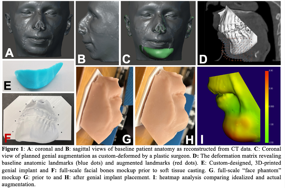
Methods: To generate a working model for protocol validation, we utilized de-identified facial CT data to provide "ground truth" patient anatomy of the maxilla and mandible of a patient with micrognathia (Weill Cornell IRB Exemption received). Using these data, 3D models of the subject's skull and soft tissue were 3D-printed with polylactic acid (PLA) on a Prusa printer (Prusa Research a.s.) and cast in silicone, respectively. This "face phantom" was imaged to generate a 3D photograph utilizing commercially available photogrammetry software (Metashape, Agisoft LLC). Desired augmentation was determined via virtual deformation of this model by a plastic surgeon. A corresponding, custom-designed genial implant was 3D-printed in PLA, implanted on the phantom, and reimaged as above. To demonstrate fidelity, the photogrammetrically-derived model with and without augmentation was co-registered and compared to CT-derived "ground truth" mesh using 3D Slicer (www.slicer.org).
Results: Model co-registration and subsequent quantitative analysis comparing the expected and actual facial deformation secondary to genial augmentation revealed a maximum Hausforff distance of 14.39 mm with an average Hausdorff distance of 1.37 mm (95% 5.22 mm; Dice coefficient=0.93). Heatmap analysis demonstrated high congruence in key relevant anatomical areas, with notable variation along the most protuberant aspect of the augmentation.
Conclusions: Our imaging protocol produces a highly accurate means of capturing critical facial anatomy necessary for design of custom-designed genial implants. We anticipate that implant fabrication customized to each patient's anatomical need will dramatically reduce cost, operative morbidity, and revisions, and produce a highly satisfactory aesthetic result. Future modeling will seek to utilize more elastic silicone to allow for a greater degree of "soft tissue" redistribution after implant placement and thus even higher aesthetic fidelity prior to translation to cadaveric models. Ultimately, we hope to generalize our protocol to other facial structures such as the nasal dorsum, cheek, and submalar region, thereby allowing for point of care production of multiple customizable facial implants.
Back to 2022 Abstracts