Hemifacial Microsomia Reconstruction Alters Layperson Visual Attention: A Prospective Study with Eye-Tracking Technology
Dillan F Villavisanis1,2, Clifford I Workman2, Zachary D Zapatero1,2, Giap H Vu1,2, Stacey A Humphries2, Jessica D Blum1, Daniel Y Cho1, Scott P Bartlett1, Jordan W Swanson1, Anjan Chatterjee2, Jesse A Taylor1
1Division of Plastic & Reconstructive Surgery, Children's Hospital of Philadelphia, Philadelphia, PA, USA1Division of Plastic & Reconstructive Surgery, Children's Hospital of Philadelphia. Philadelphia, PA, USA2Penn Center for Neuroaesthetics, Perelman School of Medicine at the University of Pennsylvania, Philadelphia, PA, USA
Background: Craniofacial and plastic surgery aim to reconstruct facial anomalies to approach “ideal normal” appearance. However, facial areas attracting the most visual attention in a variety of craniofacial pathologies are poorly understood, with a limited number of studies employing eye-tracking technology to successfully characterize these areas of interest. Hemifacial microsomia (HFM) is ideal for studying gaze patterns due to its predilection for specific facial regions, most commonly the jaws and ear. This study aimed to determine lay person visual attention toward pre- and post- jaw reconstruction of HFM patients using eye-tracking technology.
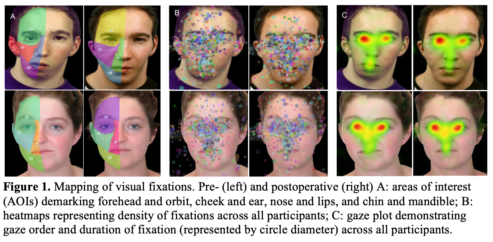
Methods: Publicly available images of seventeen patients with HFM pre- and post-jaw reconstructive procedures were used in this study. Four discrete areas of interest (AOIs) were defined on the anomalous side of each face as the forehead and orbit region, cheek and ear region, nose and lip region, and mandible and chin region. The Tobii Pro Nano eye-tracking system was used to register participant visual fixations. Each participant completed two consecutive trials consisting of 68 total images for five seconds each (34 in normal orientation and 34 in mirrored orientation to account for left gaze bias). A power analysis computed with 1,000 Monte Carlo simulations suggested a sample of 60 participants would provide sufficient power (1-β > 0.8). Linear mixed effect models (LMEMs) in R Studio tested whether locations of participant fixations were affected by surgical correction of HFM.
Results: 47,354 visual fixations were captured over 120 trials from 60 participants. LMEMs revealed significantly decreased postoperative visual fixations in the mandible and chin region [716 (54.8%) pre-reconstruction, 591 (45.2%) post reconstruction; β = -0.198, SE = 0.056, z = -3.550, p < 0.001]. Analysis also revealed significantly increased postoperative visual fixations in the forehead and orbit region [11350 (48.6%) pre-reconstruction, 12000 (51.4%) post-reconstruction; β = 0.086, SE = 0.015, z = 5.664, p < 0.00001]. Surgical reconstruction did not significantly influence visual attention to the nose and lips [8,460 (49.7%) pre-reconstruction, 8571 (50.3%) post reconstruction] or to the cheek and ears [2841 (49.9%) pre-reconstruction, 2825 (51.1%) post reconstruction] with superior fit to the null model.
Conclusions: Following corrective jaw surgery for HFM, laypersons demonstrated significantly less visual attention to the mandible and chin and increased visual attention to the forehead and orbit. These findings suggest postoperative improvement towards aesthetic normalcy may reduce visual attention toward previously anomalous anatomy.
Back to 2022 Abstracts

