Exogenous Testosterone Impairs Wound Healing in a Cross-Sex Murine Wound Model that Mimics Gender Affirming-Surgery
Erik Reiche, MD(1,2; Patrick R Keller, MD(1; Yu Tan, PhD(1; Matthew R Louis, MD(1; Vance Soares, MS(1; Calvin R Schuster, BA(1; Tingying Lu, BS(1; Vanessa Mroueh, BS(1; Devin Coon, MD, MSE(1,2
(1Department of Plastic and Reconstructive Surgery, Johns Hopkins University School of Medicine, Baltimore, MD, USA (2Division of Plastic Surgery, Brigham and Women's Hospital, Boston, MA, USA
Introduction: Wound healing problems are a major cause of morbidity for gender-affirming surgery (GAS) patients, such as hypertrophic scarring and fistula and stricture formation after phalloplasty. Prior studies have shown sex hormones may affect wound healing. To this date, however, no animal study has evaluated the effects of cross-sex hormone therapy on wound healing. We hypothesized exogenous testosterone supplementation may impair post-GAS wound healing and developed a novel murine model to investigate this phenomenon.
Methods: Mice were randomized based on hormone regimen and gonadectomy (OVX). Gonadectomy or sham occurred on day 0 and mice were assigned to no testosterone (-T), mono- or bi-weekly (T/2T) testosterone groups. Bilateral dorsal splinted wounding occurred on day 14 and wound harvest on day 21. Serum testosterone levels were quantified with mass spectrometry. Macroscopic wound healing rates were calculated through planimetry quantification of standardized wound pictures at post-operative dates 0, 3, 5 and 7. Wound tissue underwent analysis with qPCR, ELISA and immunofluorescence.
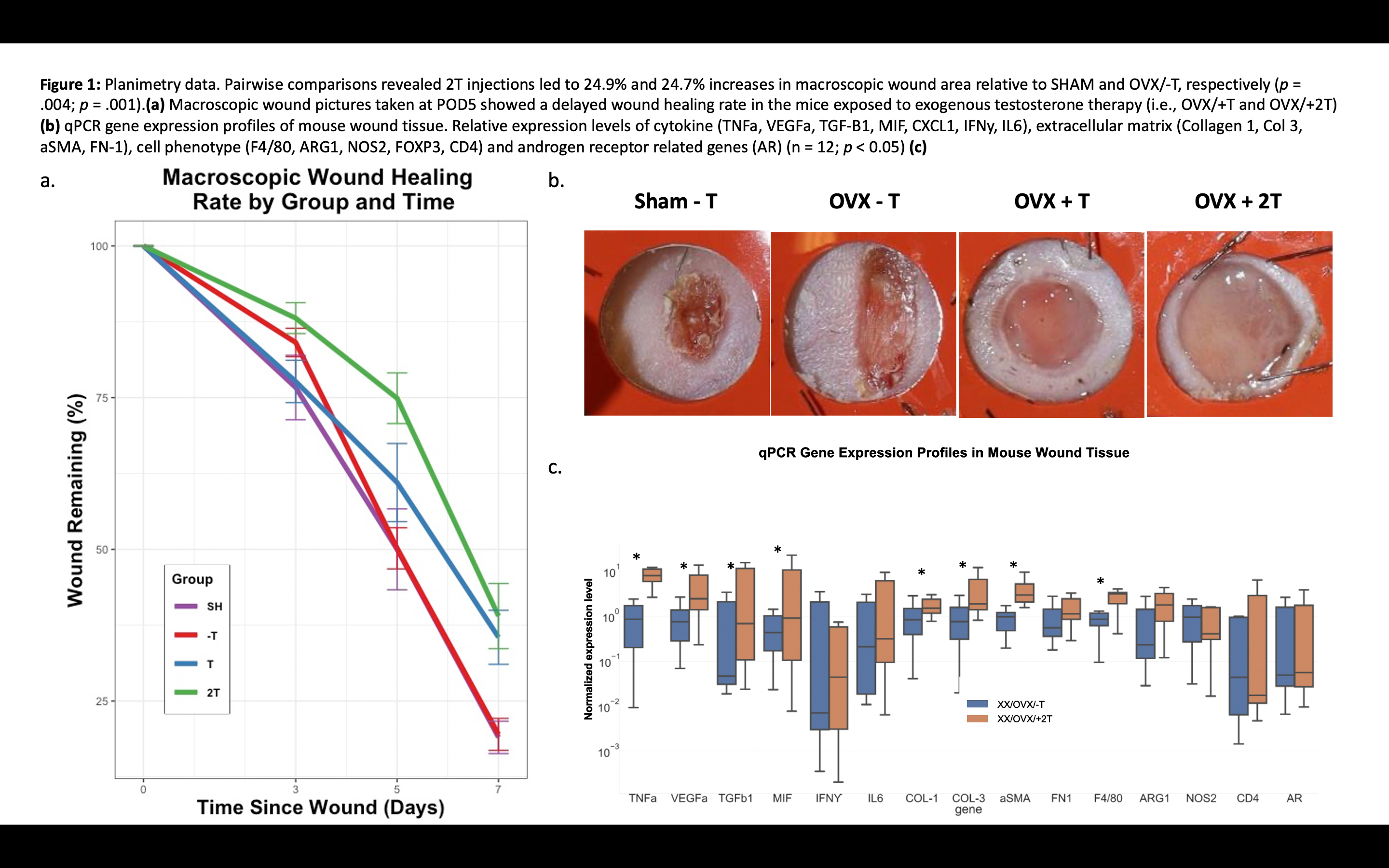
Results: Mean testosterone trough levels for bi-weekly regimen were higher compared to mono-weekly (397ng/dL vs. 272ng/dL; p=.027). At POD5, 2T injections led to 24.9% and 24.7% increases in mean wound size relative to SHAM and OVX/-T, respectively (p=.004; .001). Wounds in OVX/+2T mice demonstrated increased gene expression for inflammatory cytokines (i.e., TNFa, VEGFa, TGFb1, MIF, Col-1, Col-3, aSMA) and macrophage marker F4/80 (p<.05). ELISA confirmed elevated wound TNFa levels (p<.05). Quantitative multiplex immunofluorescence with F4/80/NOS2/ARG1 showed significant increases in macrophage prevalence in OVX/+2T (p<.05).
Discussion: We developed a novel model of the GAS hormonal milieu to study effects of exogenous testosterone on wound healing. Optimized twice-weekly dosing yielded serum levels comparable to clinical therapy. We show that exogenous testosterone administered to XX/OVX mice significantly impairs wound healing. A hyperinflammatory wound environment results with increased macrophage proliferation and elevated cytokines such as TNFa. Current efforts are directed towards mechanistic investigation and clinical validation.
Back to 2022 Abstracts

