Clinical and Histopathological Investigation of Pediatric Melanonychia
Omar Ani*1, Wen Xu2, Ines C. Lin2, Benjamin Chang2
1Perelman School of Medicine at the University of Pennsylvania, Philadelphia, PA; 2Plastic Surgery, Children's Hospital of Philadelphia, Philadelphia, PA
The natural history of pediatric melanonychia and the necessity of biopsy for ruling out melanoma are debated in the literature. We hypothesize that there is a low rate of malignant nail pathology amongst pediatric patients undergoing nail bed biopsy for melanonychia.
We performed a retrospective chart review of pediatric patients at a single institution who presented with melanonychia and underwent nail bed biopsy from 2007-2022. Univariate and multivariate analyses was performed to assess for risk factors associated with high-risk pathology findings.
The final sample included 54 patients. The average age of melanonychia onset and first biopsy was 5.5±4.4 and 7.8±4.3 years, respectively. On physical exam, 27 patients had at least four features concerning for melanoma. Pathology diagnoses included melanocytic nevus (35%), atypical intraepidermal melanocytic proliferation (AIMP) with benign features (24%), subungual lentigo (22%), and AIMP with concerning features (17%). Concerning features include increased nuclear size, nuclear atypia, and predominance of single cells over nests. There were no cases of melanoma in situ or invasive malignant melanoma. On multivariate regression, the only significant risk factor associated with more concerning pathology (AIMP with concerning features) was the calendar year in which biopsy was performed (coefficient = -0.34, p = 0.016). There was no association between physical exam features and high risk pathology.
Our case series did not find any cases of melanoma in situ or malignant melanoma rising from pediatric melanonychia. AIMP with concerning features was associated only with the year in which the biopsy was performed, which may reflect the improved understanding of pediatric melanonychia as often benign despite concerning features on pathology. Therefore, the decision to perform a nail matrix biopsy in pediatric melanonychia should be based on a collaborative discussion between the patient's parents, dermatologist, and hand surgeon.
Patient Demographics, Physical Exam, and Pathology Findings
| Average age at onset | 5.5 years | |
| Total number of patients | 54 patients | |
| Race | ||
| Male | 32 (59%) | |
| Female | 22 (41%) | |
| Asian | 12 (22%) | |
| Black | 11 (20%) | |
| Hispanic | 3 (6%) | |
| White | 27 (50%) | |
| Other | 1 (2%) | |
| Physical Exam | ||
| Asymmetry | 26 (48%) | |
| Border Irregularity | 18 (33%) | |
| Color Heterogeneity | 32 (59%) | |
| Diameter | 22 (41%) | |
| Evolving (darkening and widening) | 22 (41%) | |
| Hutchinson's Sign | 17 (31%) | |
| Pathology Results | ||
| Melanocytic Nevus | 19 (35%) | |
| AIMP with benign features | 13 (24%) | |
| Lentigo | 12 (22%) | |
| AIMP with concerning features | 9 (17%) | |
| Non-diagnostic results | 1 (2%) |

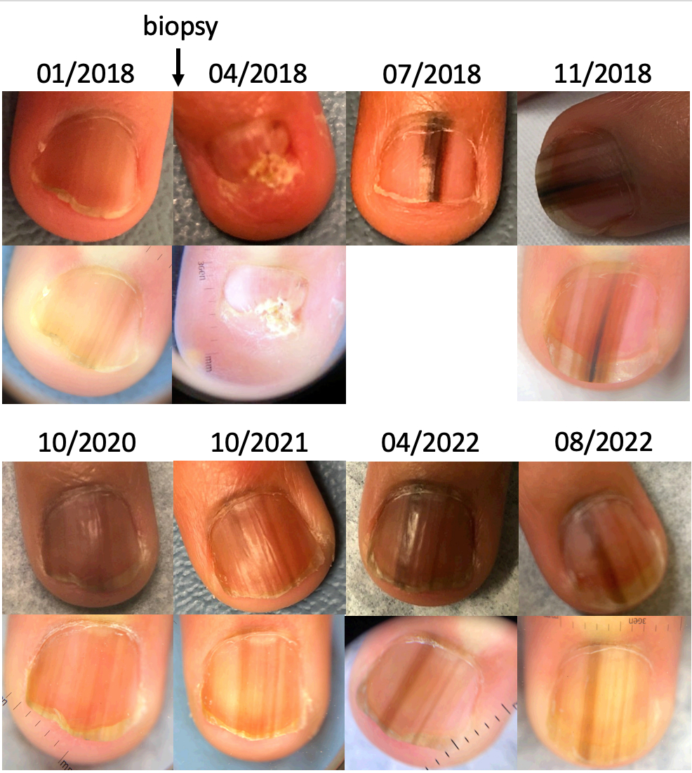
Figure 1. The course of melanonychia in a patient following biopsy exhibited alternating phases of growth and atrophy as well as increased and decreased pigmentation.
Back to 2023 Abstracts


