3D printed beta-tricalcium phosphate (β-TCP) scaffolds augmented with dipyridamole stimulate bone regeneration in an in vivo translational pediatric pig model without disruption of facial symmetry
Alexandra N. Verzella*1, Evellyn DeMitchell-Rodriguez1, Chen Shen1, Allison L. Diaz1, Andrea Torroni1, Lauren Yarholar1, Nick Tovar3, Vasudev Vivekenand Nayak3, Andre Alcon1, Jill Schechter1, Paulo Coehlo1, Bruce N. Cronstein2, Lukasz Witek3, Roberto L. Flores1
1Hansjorg Wyss Department of Plastic Surgery, NYU Grossman School of Medicine, New York, NY; 2Department of Medicine, NYU Grossman School of Medicine, New York, NY; 3Department of Biomaterials and Biomimetics, NYU College of Dentistry, New York, NY
3D printed beta-tricalcium phosphate (β -TCP) scaffolds augmented with dipyridamole (DIPY) have been shown to regenerate bone in immature small animal models. This study assesses the bone generation capacity and effects on craniofacial development of 3D printed β -TCP scaffolds + DIPY compared to autogenous bone graft using a large translational growing animal model. Assessments were made during interim growth and after full craniofacial development.
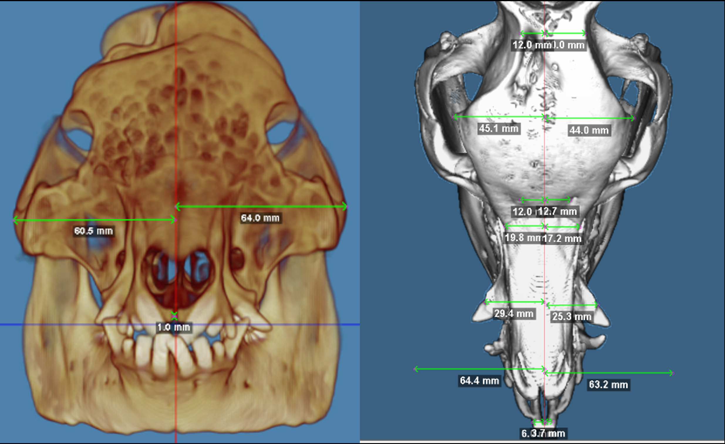
Unilateral critical-size calvarial defects and alveolar defects were created in six-week-old Göttingen minipigs (n=12). Six pigs were treated with a 3D printed β -TCP scaffolds + 1000µM DIPY, while the remaining six were treated with autologous grafts. At 12 weeks and 24 months postoperatively, the calvaria were scanned using computed tomography imaging ex vivo. Cranial symmetry, 3D-reconstruction volumetric analyses, and histological analyses were performed. Facial symmetry was evaluated using the Asymmetry Index (AI), with lower values indicating less asymmetry.
At 12 weeks, the average bone volume fraction was 84.6%, comparable to that of bone graft, and at 24 months, the 3D printed β -TCP scaffolds + 1000µM DIPY showed complete closure of defects. Histological analysis at the interim time-point demonstrated vascularized woven and lamellar bone with haversian canals. At 24-months, calvarial and alveolar defects filled with scaffolds did not demonstrate significant asymmetric growth compared to the autologous grafts when assessing global, calvarial, and alveolar right-left mediolateral asymmetry. All sutures remained patent, and there was no evidence of ectopic bone formation.
3D printed β -TCP scaffolds + 1000µM DIPY can generate bone across critical sized bone defects in a large translational animal model in a manner comparable to autogenous bone graft. All growth sutures remain patent and normal craniofacial growth appears to be preserved. Histologic analysis confirms the development of vascularized bone.
Back to 2023 Abstracts


