Use of Ultrasound in the Diagnosis of Craniosynostosis
Michael Chang*, Abigail Katz, Janet C. Coleman-Belin, Nargiz Seyidova, Olachi Oleru, Taylor Ibelli, Peter J. Taub
Icahn School of Medicine at Mount Sinai, New York, NY
Craniosynostosis (CS), caused by premature fusion of the sutures between skull bones, is a rare condition in infants that may produce increased intracranial pressure and have related detrimental effects on the growing brain. Recently, ultrasound has been suggested as a diagnostic tool for CS, although its use in clinical practice is not well characterized. The authors examined the trends of imaging for the diagnosis of CS and delineate if imaging patterns have changed with newer studies in ultrasound.
Patients with suspected CS that underwent ultrasound as their first imaging modality between January 2005 to December 2018 at Mount Sinai Hospital were evaluated. Patients that had additional scans to confirm the diagnosis were also analyzed.
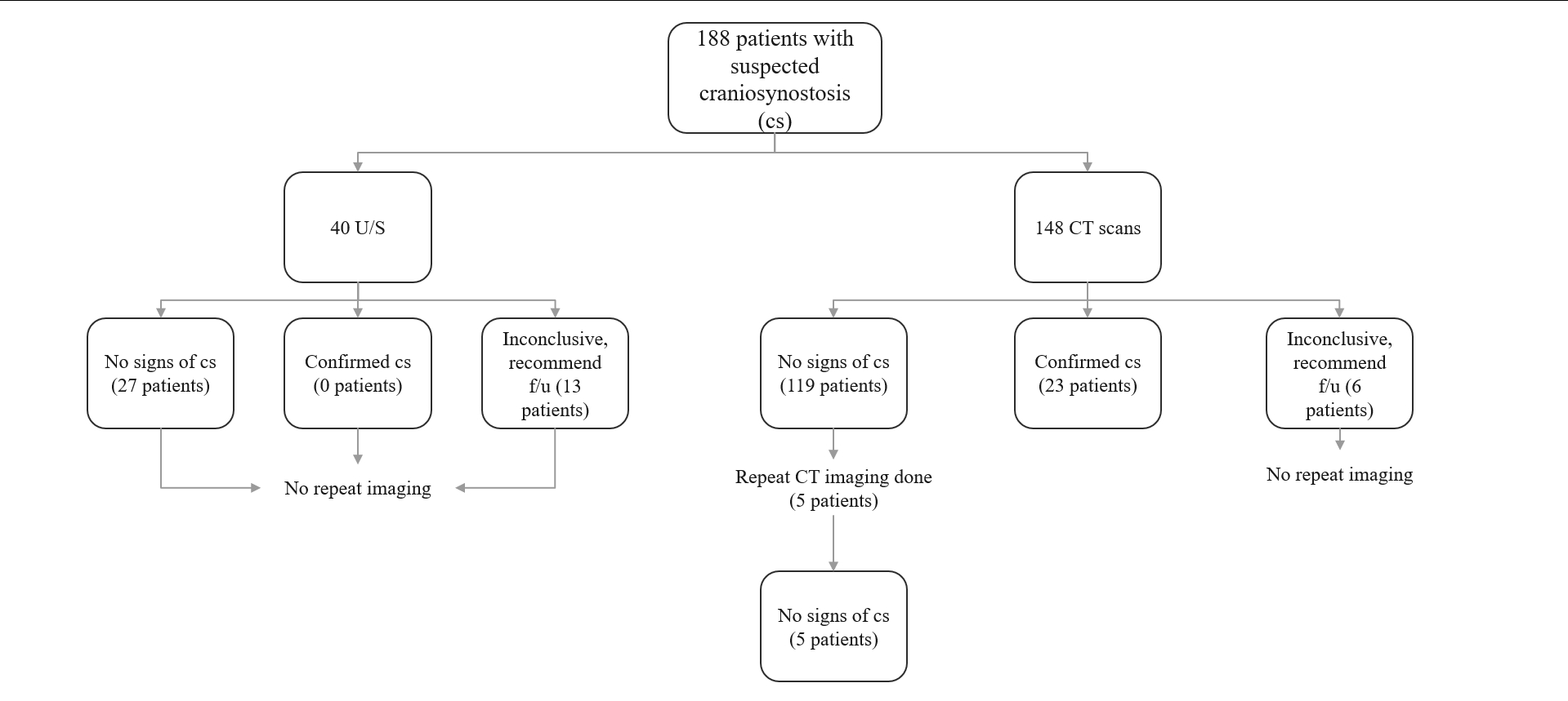
A total of 40 patients with suspected CS underwent US scans for their first imaging modality. Out of these patients, 27 did not show signs of CS, and 13 had inconclusive studies, where further imaging was not done at the discretion of the provider. No patients who initially underwent US had repeat US done. Initial CT scan for patients with suspected CS was performed in 148 patients. 23 were confirmed to have CS, 119 did not shows signs of CS, and 6 had inconclusive studies. Of the 119 CT scans that did not show signs of CS, 5 patients had repeat CT scans and all 5 had confirmed no signs of CS.
The use of ultrasound for the diagnosis of CS in a clinical setting has rarely been studied. The present study shows that many initial scans/diagnoses are made with CT. Furthermore, ultrasounds, though effective in ruling out CS, often lead to inconclusive reads that require follow-up. Physicians maintain a preference for CT imaging as the definitive diagnostic tool for CS, and they remain consistent with the imaging modality began with, when repeating scans. A potential limitation could be system-implemented protocols that result in repeated imaging modalities, such as CT followed by CT, leaving open the possibility that multiple site studies could show different imaging trends.

Distribution of imaging trends for suspsected CS at the Mount Sinai Hospital.
Back to 2023 Posters


