


Understanding the Mechanism of Radiation Damage to Human Skin
Jose A. Arellano*, Yusuf Surucu, Hamid Malekzadeh, Wayne V. Nerone, Zayaan S. Tirmizi, Rakibul Islam, Alexey Altman, Katherine Yang, Fuat Baris Bengur, Jeffrey Gusenoff, Francesco Egro, Peter Rubin, Asim Ejaz
Plastic Surgery, University of Pittsburgh, Pittsburgh, PA
Radiation-induced skin fibrosis is one of the main adverse effects of radiation therapy for cancer treatment. Radiation fibrosis syndrome is caused by the overactivation of TGF-B that promotes fibroblast that induces collagenases and breaks down type III collagen and replaces it with type I collagen. The inhibition of P53 and the accumulation of ROS are the main mechanisms of radiation-induced damage. To fully understand these mechanisms, we tested the use of our novel human skin perfusion model to recreate the damage caused by radiation.
We use our novel skin perfusion model, which consists of a human tissue sample recovered from abdominoplasty. On the first day after perfusion, we induced radiation of 20gy and 40gy to the skin and took punch biopsies on days 3, 6, 12, and 16. The samples were stained with H&E, Massons Trichrome, and measure the distance between the epidermis and the most significant collagen deposition to calculate the level of radiation-induced collagen deposition between groups, and TUNEL and DAPI to look for apoptotic changes in the epidermis/dermis.
The radiated skin started peeling compared to the control group. The H&E staining showed an increase of inflammation in the dermis along with epidermis/dermis separation as well as papillary dermis containing fibrin deposition, accumulation of inflammatory cells, reactive changes in the endothelial cells, and abundant necrotic keratinocytes. The distance of collagen deposition in the Massons Trichrome staining shows an increase in the collagen deposition in the radiation-induced skin compared to the control who had lower deposition of collagen. The TUNEL and DAPI stain shows an increase in apoptotic cells in the radiation group that correlates with the damage induced by radiation.
Our experiment was able to recreate the radiation-induced deposition of collagen shown in the H&E and Massons trichrome as well as the radiation-induced damage in the TUNEL and DAPI staining. Our system is reliable and can be used as a platform for therapeutics to mitigate the effects of radiation on the skin.
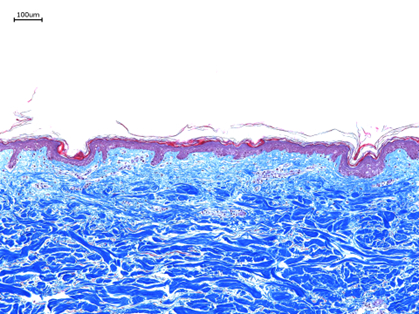
Control sample
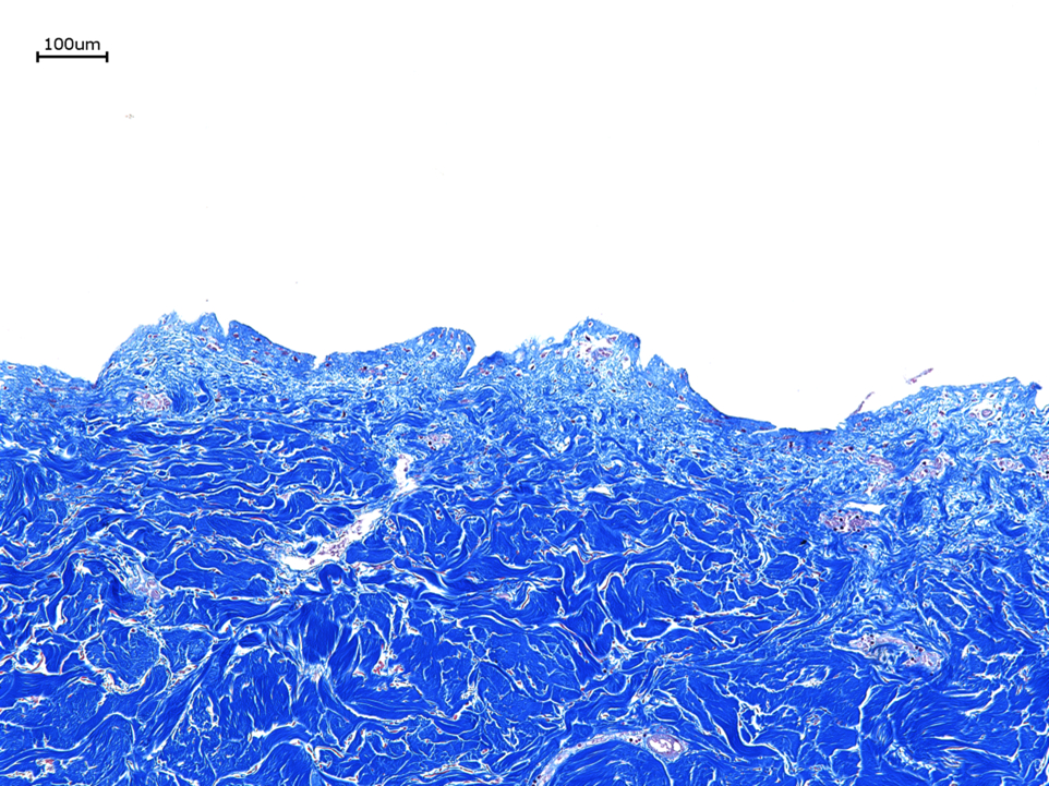
40gy radiated skin
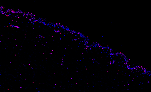
TUNEL stain showing cell death
Back to 2023 Posters