Back to 2026 Abstracts
Enhancing Patient Selection for Nerve Decompression: A standardized computerized tomography protocol for frontal and rhinogenic neuralgia
Justin Lee
1, Matthew A. DePamphilis
1, Charles D. Hwang
1, Eri Iwasaki
2, Josip Plascevic
1, Karen Buch
2, Ali Totonchi
3, William G. Austen
1, Osamu Sakai
2, Katherine Carruthers
1
1. Division of Plastic and Reconstructive Surgery, Massachusetts General Hospital, Harvard Medical School, Boston, Massachusetts, USA., Boston, MA, United States. 2. Department of Radiology, Massachusetts General Hospital, Harvard Medical School, Boston,
Background: Appropriate selection of patients with frontal or rhinogenic site neuralgia is challenging yet critical for effective surgical treatment. This study evaluates the utility of computerized tomographic (CT) imaging in preoperative assessment of frontal or rhinogenic sites and introduces a protocol to improve standardized review.
Methods: A prospective cohort study was conducted of patients undergoing frontal or rhinogenic site surgery between 2012-2025. Patients with preoperative CT imaging were included. A standardized CT review protocol was developed and applied by blinded radiologists. Radiographic findings were compared with clinical and intraoperative anatomy, and detection rates were compared with original radiology reports. Pain outcomes were assessed longitudinally.
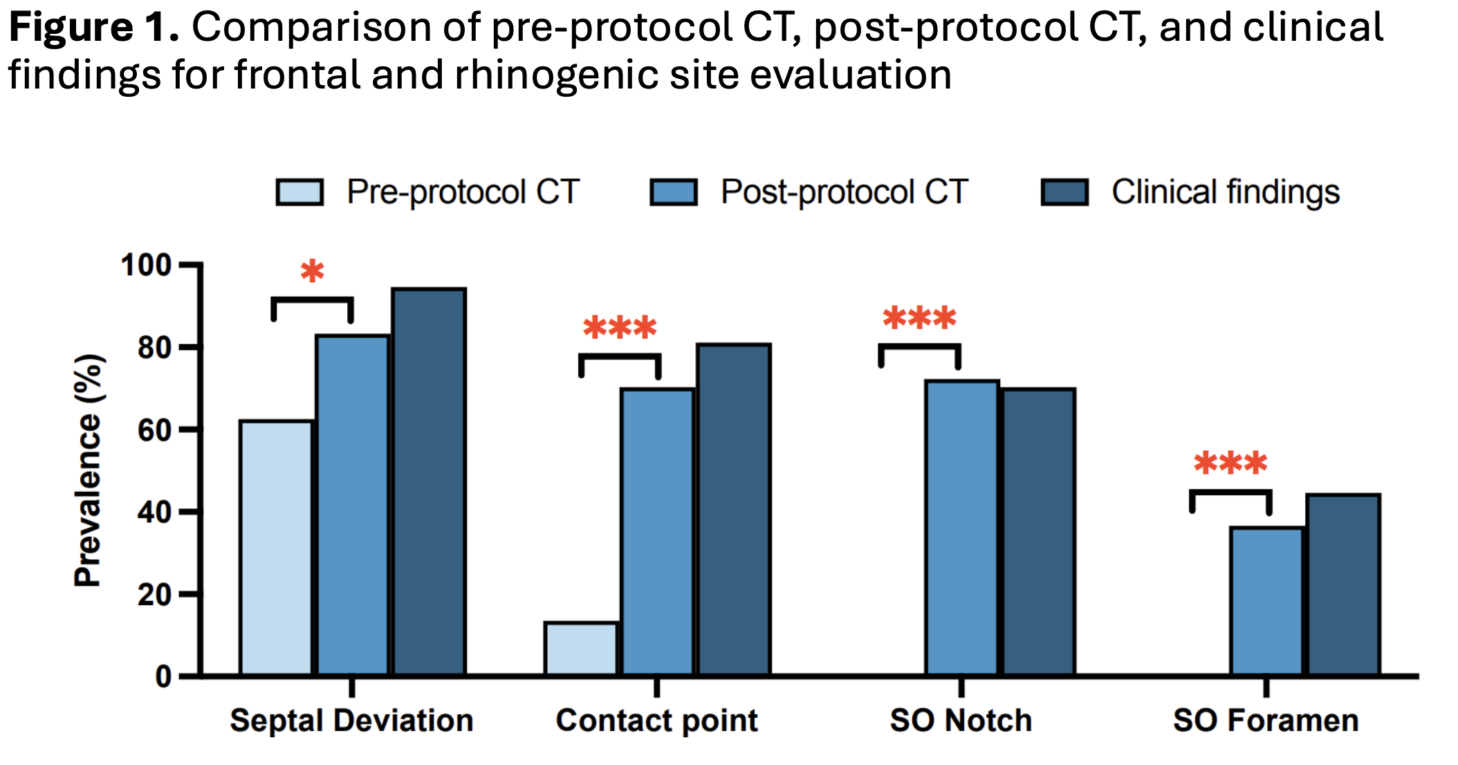
Results: Final sample included 74 patients with preoperative CT imaging. Implementation of the standardized protocol significantly improved radiographic detection of intranasal contact points (13.5% to 70.3%, p<0.001), supraorbital notch (0% to 72.3%, p<0.001), and foramen (0% to 36.6%, p<0.001). When correlated with clinical findings, CT demonstrated 83.3% accuracy for contact points, 96.0% for supraorbital notch, and 92.1% for foramen detection. Frontal decompression resulted in a median pain improvement of 75% (IQR, 50%-97%) and rhinogenic surgery improvement of 80% (IQR, 0%-100%), over mean follow-up periods of 24 and 32 months respectively.
Conclusion: CT imaging is a valuable adjunct in the preoperative evaluation of patients with suspected frontal or rhinogenic neuralgia. Relevant anatomic findings are frequently underreported in standard radiologic interpretations. Incorporation of a structured imaging review protocol into preoperative assessment may enhance patient selection, optimize surgical planning, and ultimately improve outcomes following nerve decompression surgery.
Back to 2026 Abstracts


